
13th August 2022, NIA Diagnostic Imaging
In a 2020 study, 1 in 20 Australians have diabetes mellitus (excludes gestational diabetes), and the primary complications associated with this disease are foot ulcerations and claudication (Australian Institute of Health and Welfare, 2022). Diabetic foot ulcerations can develop due to a number of factors including poor glycaemic control, poor foot care, dry skin, calluses, and foot deformities (Oliver & Mutluoglu, 2022).
However, a common cause for foot ulcerations and claudication is poor circulation due to damaging changes to nerves and blood vessels, and/or an underlying peripheral arterial disease (PAD) – primarily atherosclerosis (AT) (K.S. & Kumar A., 2020).
Risk factors of atherosclerosis:
- Age (males greater than 45-years-old and females greater than 55-years-old)
- Smoking
- Diabetes mellitus
- High lipid diet
- Sedentary lifestyle
- Family history (male family member younger than 55 years and female family member younger than 65 years)
- Systemic arterial hypertension
Pathophysiology
AT occurs when the arterial endothelium undergoes injuries (physical or from chemical toxins such as smoking) or dysfunction. These changes to the endothelial cells allow an influx of lipids to easily permeate the vessel layers and adhere to the intima wall. Intima thickening also occurs when smooth muscles cells migrate from media to intima layer. Overtime, more lipids, platelets, necrotic remnants, blood monocytes and lymphocytes, and collagen accumulate to become the visible soft and/or hard plaque seen on ultrasound (Prado dos Santos, et al., 2021).
Imaging and Diagnosis
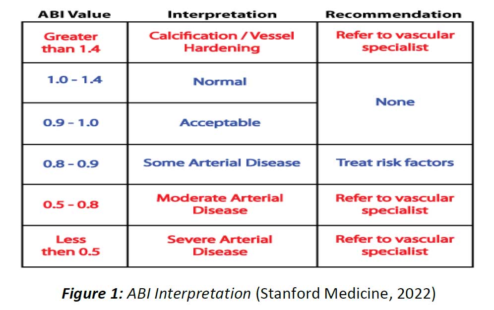
At NIA Diagnostic Imaging, arterial Doppler/Duplex studies for both upper and lower limbs are a service provided to aid in the assessment of PAD in patients – especially patients with foot ulcerations, claudication, weakness, and numbness in the limbs which are symptoms associated with diabetes mellitus. Arterial Doppler/Duplex studies involves use of colour Doppler – useful in detecting segments that appear stenotic or occlude – and pulsed wave Doppler, which detects flow velocity and degree of severity of the stenoses. The ankle-brachial index (ABI) is also performed with ultrasound guidance prior to commencing the arterial doppler/duplex study to provide an initial diagnosis.
The table below summaries interpretations of ABI values and recommended treatments associated:
Criteria
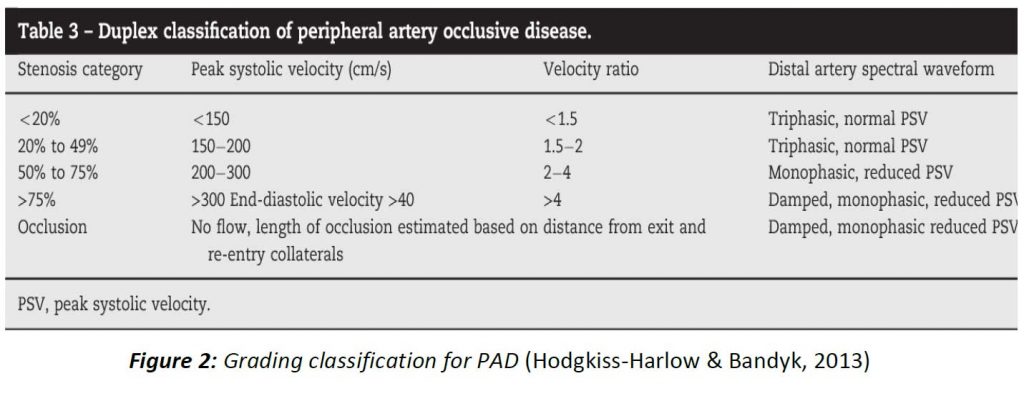
The criteria used for diagnosing occlusive disease is defined by assessing the:
– Velocity
➢ Velocity increases through stenosis lumen and can return to normal velocity post stenosis
➢ Stenosis is determined by the ratio of velocity through the stenosis lumen to the velocity proximal to the stenosis
➢ Peak velocity is greater than 200 cm/s which indicates haemodynamically significant
– Flow changes
➢ Laminar flow changes from either triphasic or biphasic to monophasic flow (Nasra & Osher, 2021)
The table below is used to help classify the degree of occlusive disease for lower limbs:
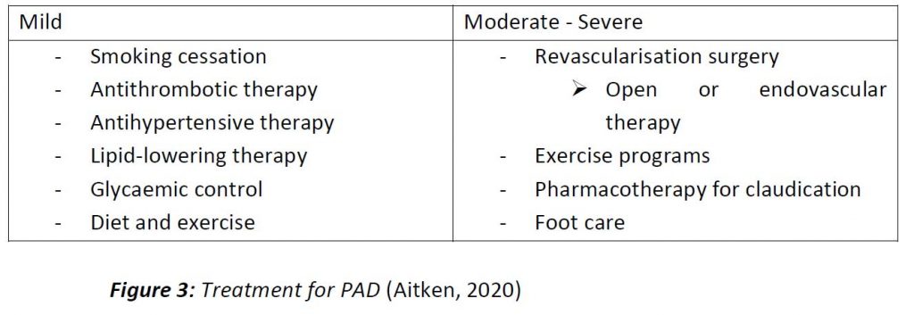
Treatment
There are various methods of treatment for PAD and the appropriate treatment is dependent on the severity of the disease.
References:
- Aitken, S., 2020. Peripheral artery disease in the lower limbs. Focus, 49(5), pp. 239-244.
- Australian Institute of Health and Welfare, 2022. Diabetes: Australian facts, s.l.: Australian Government.
- Deane, C. & Goss, D., 2011. Peripheral arteries. In: Clinical Ultrasound. England: Churchill Livingstone, pp. 1197-1226.
- Hodgkiss-Harlow, K. & Bandyk, D., 2013. Interpretation of arterial duplex testing of lower-extremity arteries and interventions. Seminars in Vascular Surgery, 26(2-3), pp. 95-104.
- Hwang, J. Y., 2017. Doppler ultrasonography of the lower extremity arteries: anatomy and scanning guidelines. Ultrasonography, 36(2), pp. 111-119.
- K.S., S. & Kumar A., S., 2020. Assessment of Diabetic Foot through the Developmental Stages of Lower Limb Abnormalities Using Ultrasound, s.l.: IntechOpen.
- Nasra, K. & Osher, M., 2021. Sonography Vascular Peripheral Arterial Assessment, Protocols, And Interpretation, Treasure Island, Florida: StatPearls.
- Oliver, T. & Mutluoglu, M., 2022. Diabetic Foot Ulcer, Treasure Island, Florida: StatPearls Publishing.
- Pahwa, R. & Jialal, I., 2021. Atherosclerosis, Treasure Island, Florida: StatPearls.
- Prado dos Santos, V., Pozzan, G., Castelli, Jr, V. & Caffaro, R. A., 2021. Arteriosclerosis, atherosclerosis, arteriolosclerosis, and Monckeberg medial calcific sclerosis: what is the difference?. Jornal Vascular Brasileiro, Volume 20, pp. 1-8.
- Stanford Medicine, 2022. Measuring and Understanding the Ankle Brachial Index (ABI). [Online] Available at: https://stanfordmedicine25.stanford.edu/the25/ankle-brachial-index.html [Accessed 11 August 2022].