
13th February 2024, NIA Diagnostic Imaging

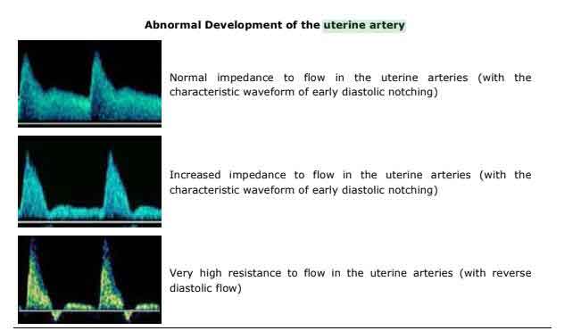
Commonly, uterine artery doppler ultrasound is conducted to measure the blood flow in the uterine arteries. This specific ultrasound allows for qualitative information to be gathered on the presence/absence of flow, the direction of flow, the quality of flow, to determine if the flow is laminar or turbulent and to distinguish waveform shape (low resistance, high resistance, early diastolic notch) (Naguib et al.,2012).
The uterine arteries are the vessels that provide blood flow to the uterus, which then supplies the placenta. There are two uterine arteries, one on the left and one on the right side of the uterus, arising from the anterior deviate of the internal iliac artery. As such, a fetus will be able to grow healthily if there is sufficient blood flow through these arteries as the placenta will develop normally (Chaudry et al.,2023).
In cases with defective trophoblast invasion, impaired placentation may occur which may result in pre-eclampsia and early onset fetal growth restriction (Chaudry et al., 2023).
At NIA Diagnostic Imaging, our highly skilled sonographers frequently perform uterine artery doppler assessment during obstetric ultrasounds, as we value the importance of assessing placental flow. Ultrasound is a highly advantageous imaging modality as it is non-invasive in nature and utilises non-ionising radiation, hence being extremely safe and beneficial for patients (Naguib et al.,2012).
At NIA Diagnostic Imaging, we utilise the latest GE LOGIQ E10 series ultrasound system to perform uterine artery doppler assessment. Assessment involves scanning both the left and right uterine arteries and reporting their PI (Pulsatility Index).
Indications of uterine artery doppler include:
• Previous history of pre-eclampsia
• Previous history of child with IUGR
• Unexplained high maternal serum AFP level
• Unexplained high HCG levels (Barati M et al., 2014).
The onset of high blood pressure after 20 weeks of pregnancy, is strongly indicative of pre-eclampsia, which severely increases the potential of adverse outcomes for both the mother and the fetus. Hence, uterine artery doppler evaluation is essential to identify pregnancies that may be at increased risk of the development of pre-eclampsia, as it often leads to severe growth restriction and small fetus for gestational age. In pregnancies complicated by intrauterine growth restriction and pre-eclampsia, the impedance to flow in the uterine arteries is increased. In these scenarios, there is an increased likelihood of:
• Shorter duration of pregnancy
• Emergency caesarean delivery
• Placental abruption
• Poorer neonatal outcome

REFERENCES
Barati M, Shahbazian N, Ahmadi L, Masihi S. Diagnostic evaluation of uterine artery Doppler sonography for the prediction of adverse pregnancy outcomes. J Res Med Sci. 2014 Jun;19(6):515-9. https://www.ncbi.nlm.nih.gov/pmc/articles/PMC4155705/#:~:text=Abnormal%20uterine%20artery%20Doppler%20result,for%20identifying%20high%2Drisk%20pregnancies.
Chaudhry, R. and Chaudhry, K. (2023) Anatomy, Abdomen and Pelvis: Uterine Arteries. https://www.ncbi.nlm.nih.gov/books/NBK482267/ Isuog (2022) Uterine arteries Doppler ultrasound, International Society of Ultrasound in Obstetrics & Gynecology. Available at: https://www.isuog.org/clinical-resources/patient-information-series/patient-information-pregnancy-conditions/growth-and-fluid/uterine-arteries-doppler-ultrasound.html (Accessed: 09 February 2024).
Naguib, N.N. et al. (2012) ‘Role of uterine artery Doppler in the management of uterine leiomyoma by arterial embolization’, Ultrasound in Obstetrics & Gynecology, 40(4), pp. 452–458. doi:10.1002/uog.11074. Nicolaides, K. et al. (2002) Doppler in Obstetrics, International Society of Ultrasound in Obstetrics & Gynecology. Available at: https://www.isuog.org/clinical-resources/patient-information-series/patient-information-pregnancy-conditions/growth-and-fluid/uterine-arteries-doppler-ultrasound.html (Accessed: 09 February 2024).