
13th March 2023, NIA Diagnostic Imaging
Over the last few weeks, we have explored the many issues relating to metabolic dysfunction associated fatty liver disease (MAFLD). We looked at what else we need to do after you diagnosed MALFD. In particular, we highlighted the need to assess the degree of fibrosis in each patient with MAFLD. With the continuous advances in technology, a wide range of new techniques and imaging modalities have emerged, thus allowing for improved diagnosis of various diseases and pathologies. Such advancements can be seen especially in the assessment and staging of liver fibrosis.
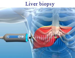
LIVER BIOPSY
According to Sande, et al. (2017), percutaneous liver biopsy is the current gold standard for the assessment and staging of liver fibrosis. In comparison to other techniques available, liver biopsy offers the lowest percentage of error (Berger, et al., 2019).
There are, however, many disadvantages that come with this procedure including:
• It is not suitable for screening purposes and early diagnosis.
• Higher risk of complications involved due to its invasive nature.
• Only a certain type of the liver parenchyma can be evaluated (Sande, et al, 2017)
FIBROSCAN
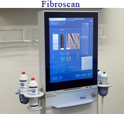
Fibroscan is a non-invasive, quick technique used to measure liver stiffness. A graphical result is displayed on the screen (Kemp & Roberts 2013). This technique is used in medical centres and hospital departments.
Disadvantages:
• Cannot be used with patients with ascites, pleural effusion, or morbidly obese patients – produces unreliable results.
• Cannot visualise areas being sampled
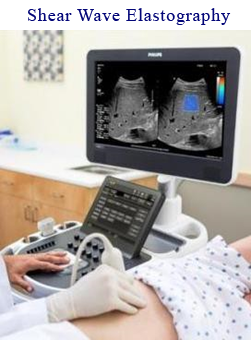
SHEAR WAVE ELASTOGRAPHY (SWE)
Shear wave elastography is a sonographic technique which evaluates the change in liver stiffness. By quantifying this stiffness, we can use this technology to assess, stage and manage liver fibrosis.
Shear wave elastography has many advantages over the previously mentioned methods:
• Non-invasive
• Low-cost
• Readily available
• Allows the examination of any area within the liver
• Can be performed easily in conjunction with a routine abdominal ultrasound scan.
• Real B-mode visualisation of the sampled areas thus avoid ascites and pleural effusion.
• Larger sample areas compared to Fibroscan which only samples at midaxillary region.
• Scan in real-time
• Has higher specificity and sensitivity than Fibroscan (Osman, shimmy & Aziz 2020).

At NIA Diagnostic Imaging, we use the latest technology with LOGIQ E10 series ultrasound system to stage fibrosis in the liver.
How shear wave elastography works
• Fibrosis is commonly graded using Metavir scores. This is acquired by sampling an area of the liver using Shear Wave Elastography and here, the stiffness of the liver parenchyma is measured and quantified.
• The acquired measurements will provide a median score in m/s units, and a Metavir score can be determined which aides in the staging of liver fibrosis.
REFERENCES
- Afdhal N. H. (2012). Fibroscan (transient elastography) for the measurement of liver fibrosis. Gastroenterology & hepatology, 8(9), 605–607.
- Berger, D., Desai, V., & Janardhan, S., Con: Liver Biopsy remains the Gold Standard to Evaluate Fibrosis in Patients With Nonalcoholic Fattu Liver Disease’, Clinical Live Disease, vol 13, issue 4, p 114-116, doi: https://doi.org/10.1002/cld.740
- Kemp, W., Roberts, S., (2013) ‘FibroScan and transient elastography’, Australian Family Physician, vol 42, issue 7, https://www.racgp.org.au/afp/2013/july/fibroscan
- Osman, A. M., El Shimy, A., & Abd El Aziz, M. M. (2020). 2D shear wave elastography (SWE) performance versus vibration-controlled transient elastography (VCTE/fibroscan) in the assessment of liver stiffness in chronic hepatitis. Insights into imaging, 11(1), 38. https://doi.org/10.1186/s13244-020-0839-y
- Sande, J, Verjee, S, Vinayak, S, Amersi F & Gheani, M 2017, ‘Ultrasound shear wave elastography and liver fibrosis: A Prospective Multicentre Study’, World Journal of Hepatology, vol 9, no. 1, pp. 38-47, doi: 10.4254/wjh.v9.i1.38
- https://ge-ultrasound.eu/wp-content/uploads/2021/02/1.-LOGIQ-E9-LOGIQ-E10-LOGIQ-E10s_Shear-Wave-Whitepaper_2020_JB29031XX.pdf
- https://gpvoice.com.au/index.php/2022/06/12/nafld-the-many-treatment-options-in-the-pipeline/
- https://gpvoice.com.au/index.php/2018/01/31/non-invasive-prenatal-testing-what-gps-need-to-know/