
11th October, 2023, NIA

Deep vein thrombosis (DVT) is a medical condition associated with significant morbidity and mortality. It is vital that a rapid diagnosis can be acquired since DVT can result in severe complications including the development of pulmonary embolism (PE). The consequences of PE can be life-threatening. About 10% of PEs are fatal and 5% will cause death later despite diagnosis and treatment.
Ultrasound is the imaging modality of choice when assessing DVT due to its many advantages including being safe, non-invasive, low-cost and reliable. A rapid diagnosis can also be made which is vital to ensure timely treatment and management can be undertaken when necessary.
At NIA Diagnostic imaging, we employ the GE LOGIC E10 ultrasound system to effectively interrogate the veins from the common femoral vein to the distal calf veins when ruling out the presence of a DVT. The patient’s affected leg is examined and compressed by a linear ultrasound probe to ensure that the veins are completely compressible. Compression of the vein on B-mode imaging can show the vessel collapsing in real-time thus, proving that no obvious DVT is observed. Non-compressibility of the vein, however, is an indicator of the presence of a DVT (Schick and Pacifico. 2022).
Another criterion to diagnosis DVT on ultrasound is the direct visualisation of the thrombus within the lumen of the vessel. The thrombus on grey-scale imaging may demonstrate variable echogenicity hence is noncompressible and may generally dilate the lumen in the acute phases of the DVT (Sosthene et al. 2019).
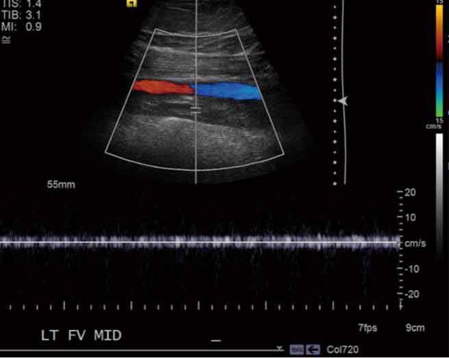
Ultrasound can also be effective in demonstrating the occlusion at the site of the thrombus by employing colour and spectral Doppler imaging. The colour Doppler image will reveal an absence or decreased flow within the lumen of the vein and a lack of a significant waveform may be visualised on the spectral Doppler trace. A loss of venous flow augmentation may also be observed with dynamic testing. (Stone et al. 2017).
Therefore, ultrasound can effectively provide a rapid diagnosis of DVT, which allows prompt treatment and management for the prevention of PE.
NIA Diagnostic Imaging understands the importance of early DVT diagnosis and is always willing to accept urgent requests for venous doppler exams. For more information, please contact:
NIA Glenquarie Radiology (02) 9158 8660 info@niaimaging.com.au
NIA Ingleburn Radiology (02) 8104 0803 info.ingleburn@niaimaging.com.au

References
1. Schick M and Pacifico L (2022) Deep Venous Thrombosis of the Lower Extremity, StatPearls, accessed on 17 September 2023. https://www.ncbi.nlm.nih.gov/books/NBK470381/
2. Sosthene T, Serge K, Medard K, Simplice K, John M and Albin S (2019) ‘Duplex ultrasound in upper and lower limb deep venous thrombosis’, Annals of Circulation, 5(1):001-007, doi: 10.17352/ac.000015
3. Stone J, Hangge P, Albadawi H, Wallace A, Shamoun F, Knuttien M, Naidu S and Oklu R (2017) ‘Deep vein thrombosis: pathogenesis, diagnosis and medical management’, Cardiovascular Diagnosis and Therapy, 7(3):276-284, doi: 10.21037/cdt.2017.09.01
4. Turetz M, Sideris A, Friedman O, Triphathi N and Horowitz J (2018) ‘Epidemiology, pathophysiology and natural history of pulmonary embolism’, Seminars in Interventional Radiology, 35(2):92-98, doi: 10.1055/s-0038-1642036