28th September 2024, A/Prof Chee L Khoo

Patients are often given tacit recommendations about lifestyle changes for MAFLD because it’s “just a little fat in the liver”. For those of us who have that few patients with those liver as well as non-liver complications, we will remember how horrible these patients fare moving forward. Yet, we can’t refer all our patients with fatty liver to the hepatologist. Most of these patients can be safely and appropriately managed and monitored in primary care. The Gastroenterological Society of Australia (GESA) has just released the latest consensus statement on assessment of MAFLD (1). GPs also have access to a series of incredibly comprehensive and practical case based online learning module to keep us uptodate. More of that later.
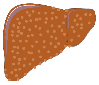
Metabolic dysfunction-associated fatty liver disease (MAFLD) is sometimes known as metabolic dysfunction-associated steatotic liver disease (MASLD) for those who think the word “fatty” is stigmatising.In addition, MASLD the definition of MASLD requires exclusion of excessive alcohol consumption (defined as ≥20 g/day for women and ≥30 g/day for men) and alternative forms of liver disease. MAFLD replaces the old term non-alcoholic fatty liver disease (NAFLD) and is embedded in the new consensus definition of steatotic liver. It should be thought of in patients with type 2 diabetes (T2D), obesity or ≥ 2 metabolic risk factors (hypertension, dyslipidaemia, prediabetes).
MAFLD is an increasingly frequent cause of cirrhosis and hepatocellular carcinoma. With obesity and its associated metabolic derangements rapidly increasing, the incident cases of advanced liver disease and liverrelated deaths due to MAFLD are estimated to increase by 85% in Australia between 2019 and 2030 (2). Most of these patients have frequent interactions with their GPs but often the prevalence of MAFLD is underappreciated and hence, underdiagnosed. Even when diagnosed, it is often not taken seriously. The consensus statement is targeted at primary care although health care professionals in other specialties will also benefit.
Diagnosis – who to screen
Most epidemiological studies using the MAFLD definition have been conducted in Asia, where the prevalence varies between and within countries:
China 21%–47%
Hong Kong 26%
South Korea 34%–38%
Japan 35% and
Sri Lanka 33% (3)
In comparison, studies from North America report prevalences between 20% and 38%, a study from the Netherlands in an older adult population reporting a MAFLD prevalence of 34%.
The diagnostic criteria pretty much says it all in relation to who is likely to have MAFLD. A number of meta-analyses in Australia found the prevalence of MAFLD amongst patients with T2D is between 54-58%. This will be a high risk group to screen for MAFLD.
The prevalence of MAFLD amongst patients with obesity ranged from 55-75%. Another way of looking at the prevalence is to look at the relative risks of MAFLD amongst patients with obesity. It ranges between 2.67 to 32-fold depending on the population studied. This will be another group which is likely to have MAFLD.
Of note, patients who have at least 2 metabolic risk factors (See Table 1) have 2.4-9.6X risk of having MAFLD even amongst those with normal weight. Thus, patients with normal weight can have MAFLD too.
What tests to order
Modelling studies done overseas have questioned the cost-effectiveness of liver ultrasound as a screening tool. Generalisation of those studies to the Australian setting is low. Liver ultrasound is a widely studied, freely available and relatively inexpensive imaging modality to detect hepatic steatosis in primary care. We should be aware of the lower sensitivity of ultrasound in patients with obesity or a low degree of hepatic steatosis.
Controlled attenuation parameter (CAP) using Fibroscan® and MRI are not readily accessible in the community and do not attract an MBS rebate for diagnosis of hepatic steatosis.
Recommendation
Liver ultrasound should be the first-line test to diagnose hepatic steatosis in people at high risk of MAFLD.
Biomarkers?
Several non-commercial biomarkers have been evaluated to identify or detect MAFLD at a population level, including the FLI, hepatic steatosis index, MAFLD liver fat score, visceral adiposity index and triglyceride 9 glucose index. These are not well suited to screening for MAFLD in primary care because of the multiple components required and the complex algorithms involved. Commercial biomarker tests for hepatic steatosis are expensive and not readily available in Australia.
Assessment for fibrosis
MAFLD may cause liver injury and inflammation (known as steatohepatitis) and can lead to liver fibrosis. Liver fibrosis is graded from zero (no fibrosis) to one (mild fibrosis), two (significant fibrosis), three (advanced fibrosis) and four (equivalent to cirrhosis). The gold standard for grading is liver biopsy although we now have non-invasive tests to quantify the fibrosis. Early identification of people at higher risk of adverse liver-related outcomes provides an opportunity for intervention to reduce disease progression and enable referral for specialist hepatology care before liver related complications develop.
Your standard LFTs are NOT sufficient in detecting advanced liver fibrosis and may even give normal results in the presence of cirrhosis. Similarly, ultrasound and CT are inaccurate for determining advanced fibrosis and lack sensitivity for determining cirrhosis.
Elastography methods assessing physical properties of the liver (e.g. liver stiffness) are sometimes utilised to assess liver fibrosis. In primary care where the prevalence of advanced fibrosis is low, the negative predictive value (NPV) is high and positive predictive value is low.
The FIB-4 Index is a non-commercial, low-cost, non-invasive test using common laboratory test results that are widely available in clinical practice: AST, ALT and Platelet Count. FIB-4 index can be easily performed with online calculators. You also have to enter the age of the patient in the calculation.
A lower FIB-4 score cut-off of 1.3 is 74% sensitive for the diagnosis of advanced fibrosis, whereas a cut-off of 2.67 is 94% specific (4). Results between these cut-offs are indeterminate. In an Australian cohort of 628 patients with MAFLD referred from primary care, the likelihood of those with a low FIB-4 score (<1.3) developing HCC or hepatic decompensation was 1% or lower over a median follow-up of 5 years (5).
Age affects the accuracy of the FIB-4 Index. FIB-4 is inaccurate in people younger than 35 years (6) although the risk of advanced liver fibrosis in young adults is very low. The specificity of FIB-4 also reduces with increasing age, such that an upper cut-off score of 2.0 is recommended.
FibroScan®, quantifies the elasticity of the liver parenchyma by sonographically measuring the velocity of an impulse induced by a mechanical driver held next to the patient’s abdominal wall. It’s recommended for patients older than 65 years. However, Fibroscan* is not Medicare rebatable and there is limited availability across Australia.
Shear wave elastography (SWE) also assesses the stiffness of the liver by quantifying “shear wave” velocity or tissue displacement generated by an ultrasonic impulse. SWE is less validated than Fibroscan®. Accuracy is very experience dependent.
Two direct serum fibrosis tests have limited availability in Australia: the ELF test and Hepascore.
Recommendations
People with MAFLD and a FIB-4 score >2.7 or elevated results of a direct liver fibrosis serum test or liver elastogram should be referred to a clinician with expertise in liver disease
A second-line assessment with liver elastography or a direct liver fibrosis serum test should be performed in people with MAFLD and a FIB-4 score between 1.3 and 2.7. If these are unavailable, referral to a clinician with expertise in liver disease should be considered.
In summary, the prevalence of MAFLD is high especially in patients with T2D, obesity and patients with 2 or more metabolic risks. MAFLD can also be present in patients who are normal weight and in patients with normal LFTs. The first line investigation to diagnose MAFLD is plain old liver ultrasound.
Once MAFLD is diagnosed, we need to assess for liver fibrosis. The FIB-4 score is well validated and is easy to use. Patients with low FIB-4 score (<1.3) is unlikely to have advanced fibrosis and safely be managed in primary care. Patients with high FIB-4 score (>2.7) should be referred to a clinician with expertise in liver disease. Patients with intermediate score should have either Fibroscan®* or shear wave elastography if Fibroscan® is not available. Patients with advanced fibrosis should be referred.
In the next issue, we will cover monitoring of progression of fibrosis in primary care, surveillance for hepatocellular carcinoma and assessment of patients with MAFLD and other causes of liver disease.
You can get access to the free online educational program here,

References:
- https://www.gesa.org.au/public/13/files/Education%20%26%20Resources/Clinical%20Practice%20Resources/MAFLD/MAFLD%20consensus%20statement%202024.pdf
- Adams LA, Roberts SK, Strasser SI, et al. Nonalcoholic fatty liver disease burden: Australia, 2019–2030. J Gastroenterol Hepatol 2020; 35: 1628-1635.
- Vaz K, Clayton-Chubb D, Majeed A, et al. Current understanding and future perspectives on the impact of changing NAFLD to MAFLD on global epidemiology and clinical outcomes. Hepatol Int 2023; 17: 1082-1097.
- Mozes FE, Lee JA, Selvaraj EA, et al. Diagnostic accuracy of non-invasive tests for advanced fibrosis in patients with NAFLD: an individual patient data meta-analysis. Gut 2022; 71: 1006-1019.
- Wang Z, Bertot LC, Jeffrey GP, et al. Serum fibrosis tests guide prognosis in metabolic dysfunction-associated fatty liver disease patients referred from primary care. Clin Gastroenterol Hepatol 2022; 20: 2041-2049.e5.
- McPherson S, Hardy T, Dufour JF, et al. Age as a confounding factor for the accurate non-invasive diagnosis of advanced NAFLD fibrosis. Am J Gastroenterol 2017; 112: 740-751.
