13th October 2024, NIA Diagnostic Imaging

Menopause is characterised by complete absence of menstrual cycle due to no ovarian follicles left in reserve and it is clinically diagnosed when a woman has amenorrhea for at least 1 year (Sung, 2023).
Any post-menopausal women with vaginal bleeding should be appropriately managed through comprehensive clinical examinations and diagnostic studies; these include diagnostic imaging and, in some cases, endometrial biopsies (Sung, 2023).
Some common causes of post-menopausal bleeding include:
● Endometrial hyperplasia
● Uterine fibroids
● Endometrial polyps
● Urogenital infections e.g. vaginitis, cystitis, endometrial tuberculosis or cervicitis
● Genitourinary atrophy
● Vaginal or endometrial atrophy (Sung, 2023).
One of the most common causes of post-menopausal bleeding is atrophy of the vagina or endometrium due to decreased estrogen and it is the presenting symptom for nearly 90% of women with endometrial cancer therefore post-menopausal bleeding is a common indication for ultrasound evaluation. It is very important to quickly and accurately diagnose or rule out neoplasia in patients presenting with this issue (Buckley & Kondagari, 2023). Ultrasound images showing thin endometrial lining (less than or equal to 4mm) has a greater than 99% negative predictive value for endometrial cancer (ACOG, 2018).

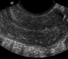
Figure 1. 39-year-old premenopausal woman with normal endometrium immediately following menses. Transvaginal sagittal ultrasound shows endometrium as a thin echogenic line that measures 3 mm (Sadro, 2016).
Endometrial hyperplasia covers pathological changes in the uterine glands and stroma. Hyperplasia can be simple or complex, with or without atypia. The presence of atypia is the most worrisome feature (Brand, 2007).
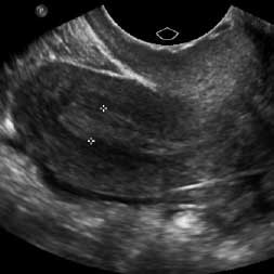
Appearance on ultrasound can include asymmetrical focal thickening with surface irregularity, heterogeneous, polypoid mass lesion, intrauterine fluid collection and myometrial invasion.

Figure 2. A 66-year-old woman with postmenopausal bleeding. Transvaginal sagittal ultrasound shows thickened heterogeneous endometrium measuring 6 mm (Sadro, 2016).
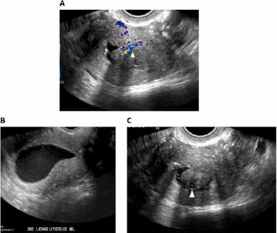
Endometrial polyps are estrogen sensitive and their incidence declines after menopause (Brand, 2007). They are associated with Tamoxifen use and appearance on ultrasound can include solitary homogenous and echogenic lesions, a stalk to the polyp with vascularity, bright edge sign and may be surrounded by endometrial fluid.

Figure 3. Transvaginal ultrasound with colour Doppler of the uterus demonstrates an endometrial mass with feeding vessels (arrowhead) consistent with an endometrial polyp (Hurtado & Mahesh,2017)
Ultrasound is the first imaging modality of choice for the female pelvis. It allows for real time, fast and portable imaging of the uterus, cervix, ovaries and adnexa without the use of ionising radiation (Buckley & Kondagari, 2023). At NIA Diagnostic Imaging, transabdominal and transvaginal ultrasound is used to measure and assess endometrial thickness and it is often recommended as a preliminary ‘non-invasive’ technique for assessing the endometrium in women with post-menopausal bleeding.
References
Buckley, E., & Kondagari, L. (2023, January 16). Sonography postmenopausal assessment, protocols, and interpretation. StatPearls [Internet]. https://www.ncbi.nlm.nih.gov/books/NBK570641/#:~:text=Postmenopausal%20bleeding%20is%20a%20common,of%20women%20with%20endometrial%20cancer
Brand, A. (2007, March). The woman with postmenopausal bleeding. https://www.racgp.org.au/getattachment/f759bd34-1266-4d94-bf7c-4e80ce0c788f/attachment.aspx
Hurtado, S., & Mahesh, S. (2017). Endometrial polyp. Endometrial Polyp – an overview | ScienceDirect Topics. https://www.sciencedirect.com/topics/medicine-and-dentistry/endometrial-polyp
Sung, S. (2023, December 20). Postmenopausal bleeding. StatPearls [Internet]. https://www.ncbi.nlm.nih.gov/books/NBK562188/
Sadro, C. (2016, April 15). Imaging the endometrium: A pictorial essay. Canadian Association of Radiologists Journal. https://www.sciencedirect.com/science/article/pii/S0846537115001345
The role of transvaginal ultrasonography in evaluating the endometrium of women with postmenopausal bleeding. ACOG. (2018, May). https://www.acog.org/clinical/clinical-guidance/committee-opinion/articles/2018/05/the-role-of-transvaginal-ultrasonography-in-evaluating-the-endometrium-of-women-with-postmenopausal-bleeding
