
12th April 2025, NIA Diagnostic Imaging
Uterine fibroids, also referred to as leiomyomas, are the most prevalent benign tumors in women of reproductive age, originating from smooth muscle cells in the uterus (myometrium) (1). While benign, uterine fibroids can greatly affect a woman’s daily quality of life (4). Ultrasound is the first-line modality used for diagnosing and characterising these fibroids (5). As GPs, we are the first port of call in the diagnosis and management of this common condition.
What are the signs and symptoms? Fibroids can either present as an asymptomatic incidental finding on imaging or symptomatically (1). Common signs and symptoms include (1, 4):
- Abnormal uterine bleeding
- Heavy or prolonged menstrual bleeding
- Pelvic pain
- Reproductive failure
- Pelvic bulk symptoms
What is the first diagnostic tool for uterine fibroids?
Transvaginal ultrasound (TVUS) is the first imaging tool recommended for the diagnosis and characterisation of uterine fibroids(4) due to its widespread availability, ease of use, and high sensitivity (90-99%) and specificity (1, 2). The purpose of TVUS is to identify the presence of fibroids, assess the size and location of each lesion, and to assess their nature (2).
What are the types of fibroids?
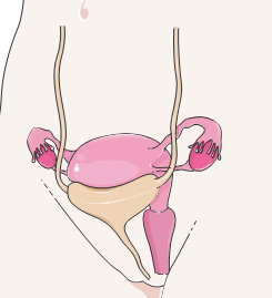
- Fibroids are typically seen in three locations (1):
- Subserosal (on the outer surface of the uterus)Intramural (inside the myometrium)
- Submucosal (inside the uterine cavity)
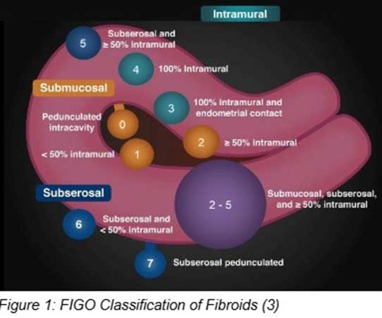
It can be further broken down into pedunculated or not. The International Federation Of Gynecology and Obstetrics (FIGO) developed a system for describing and classifying fibroids (2). What are the clinical management and treatment options for uterine fibroids? Clinical management and treatment planning are individualized and depend on several factors, including the patient’s age, symptom severity, comorbidities, characteristics of the nodules, and the desire for fertility preservation (4, 5).For women with asymptomatic fibroids, surveillance is typically recommended. Medical management, which includes hormonal contraceptives, GnRH agonists (e.g., leuprolide), and NSAIDs, aims to reduce the severity of bleeding and pain. Surgical options may involve uterine artery embolization, myomectomy, or hysterectomy (1).

References
Barjon, K., & Mikhail, L. N. (2023). Uterine leiomyomata. StatPearls Publishing. https://www.ncbi.nlm.nih.gov/books/NBK546680/ Centini, G., Cannoni, A., Ginetti, A., Colombi, I., Giorgi, M., Schettini, G., Martire, F. G., Lazzeri, L., & Zupi, E. (2024). Tailoring the diagnostic pathway for medical and surgical treatment of uterine fibroids: A narrative review. Diagnostics, 14(18), 2046. https://doi.org/10.3390/diagnostics14182046
Knipe, H. (2022). FIGO classification system for uterine leiomyoma. [Image]. Radiopaedia. https://doi.org/10.53347/rID-156167 Mension, E., Calaf, J., Chapron, C., Dolmans, M. M., Donnez, J., Marcellin, L., Petraglia, F., Vannuccini, S., & Carmona, F. (2024). An update on the management of uterine fibroids: Personalized medicine or guidelines? Journal of Endometriosis and Uterine Disorders, 7. https://doi.org/10.1016/j.jeud.2024.100080
Palheta, M. S., Medeiros, F. D. C., & Severiano, A. R. G. (2023). Reporting of uterine fibroids on ultrasound examinations: An illustrated report template focused on surgical planning. Radiologia Brasileira, 56(2), 86–94. https://doi.org/10.1590/0100- 3984.2022.0048