
25th October 2026, A/Prof Chee L Khoo

We all know that hypertension, hyperglycaemia, obesity and elevate LDL-C are risk factors for coronary artery disease. We have also seen that elevated lipoprotein (a) and elevated triglycerides are independent risk factors. We are aware that patients with systemic inflammation (e.g. psoriasis, systemic lupus erythematosus, inflammatory bowel disease, obesity, type 2 diabetes, and non-obstructive coronary syndromes) are also at higher CV risk. Biomarkers of inflammation like hsCRP are useful to gauge inflammation but agents that target inflammatory cytokines either have not been shown to reduce CV events or have shown to reduce CV events without reducing those biomarkers. Do we have a better marker of inflammation which can better quantify the CV risk?
Perivascular adipose tissue (PVAT) is the layer fat around arteries. It was once considered a harmless structural layer but is now viewed as an active participant in vascular health [1-5]. It is a heterogeneous tissue composed not only of adipocytes but also of fibroblasts, mesenchymal stem cells, macrophages, eosinophils and lymphocytes. PVAT around different vascular beds have distinct adipocyte morphology, precursor origin, secretory profile, and inflammatory potential [6]. For example, thoracic PVAT appears protective (anti-inflammatory, anti-atherogenic in mice), whereas abdominal PVAT, mesenteric PVAT, and coronary PVAT (PCAT) are associated with pro inflammatory environments and atherosclerosis susceptibility [7].
The development of atherosclerosis follows the “inside out” model, whereby atherosclerosis begins within the arterial wall due to endothelial dysfunction, inflammation, and foam cell formation. Recent research painted a more “outside” in” theory whereby dysfunctional PVAT acts as a responsive sensor to inflammatory stimuli and communicates bi-directionally with the arterial wall [3].
Adiponectin has potent anti-inflammatory and antioxidant properties and promotes vasodilation [8]. Normally (in other perivascular tissues), oxidative stress leads to lower amounts of adiponectin and endothelial dysfunction. In the PCAT (i.e coronary PVAT), oxidative stress cause adiponectin gene expression leading to increased inflammatory response with increased production of pro-inflammatory substances such as leptin, IL-6, TNF-α, IFN-γ, and monocyte chemoattractant protein-1 (MCP-1), which diffuse into the adjacent PVAT, further fueling the inflammatory response and ultimately leading to vascular dysfunction and an increased risk of developing CVD [9,10].
Zhang et al. and Dai et al., have reported no significant correlation between FAI and markers such as hs-CRP or the absolute neutrophil count [11,12].
PVAT dysfunction may also contribute to the development of hyperglycaemia and type 2 diabetes [13]. Obesity profoundly alters the phenotype and function of PVAT, promoting a shift toward larger, lipid-rich white adipocytes and a pro-inflammatory secretory profile, driving vascular dysfunction from the outside in.
FAI (fat attenuation index) is a computed tomography-derived marker of perivascular inflammation and has emerged as a novel imaging biomarker to quantify systemic or local inflammation (9). FAI values can vary between different coronary artery segment. FAI measured in the proximal right coronary artery (RCA) and left anterior descending artery (LAD) independently predicted both all-cause and cardiac mortality, whereas left circumflex artery (LCx)-derived values were less prognostic or pointed to different endpoints [14].
Vascular inflammation causes the composition of PVAT to change from a lipid-rich state to a more aqueous state. This shift leads to an increase in the density (attenuation) of PVAT on CT scans [3]. Thus, FAI can quantify coronary inflammation by plotting the attenuation gradients in peri-coronary fat. vascular inflammation can induce changes in adjacent PCAT, leading to lipolysis and a reduction in lipid accumulation. This shift results in a higher water-to-fat ratio, causing the HU values to become less negative (closer to −30 HU). Consequently, a higher FAI often indicates a greater level of coronary inflammation.
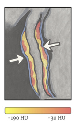
Adapted from Nˇemeˇcková, J Clin Med, July 2025
AI-corrected FAI-Score now automatically segments the coronary tree, measures three-dimensional attenuation gradients, and then adjusts for scanner voltage, age, sex, and artery-specific anatomy, outputting a unit-free score that is interpreted against age- and sex-stratified nomograms, enabling physicians to incorporate FAI into personalized prevention and therapeutic strategies.
Does FAI help predict future CV event?
In the CRISP-CT study 4000 consecutive patients with indications for CTCA had their anonymised CTCA images undergo mapping for FAI by independent investigators who were blinded to the patient demographics and outcomes (14). Perivascular fat attenuation mapping was done around the three major coronary arteries—the proximal right coronary artery, the left anterior descending artery, and the left circumflex artery. The were two separate cohort of patients. Median follow up was between 54-72 months.
The FAI values around the RCA and LAD proximal segment but not around the LCx were predictive of all-cause and cardiac mortality. They used −70.1 HU as the optimal FAI cut-off value. Values above this threshold were associated with a 9X increase in cardiac mortality, 2.5X for all-cause mortality and a 5X increased risk of acute myocardial infarction. The predictive value of the FAI for cardiac mortality was lost in those who started taking statins and aspirin after CCTA, whereas among those who did not change their medication, the hazard ratio for cardiac mortality more than doubled.
Now, we know that high dose statins has been shown to change the composition of intraplaque components by increasing calcification and decreasing the amount of non-calcified plaque burden (15). The link between FAI and CV risks was further confirmed by Matyas et al in a small study (16). They used the CaRi-Heart® system (an AI imaging solution) to quantify the degree of inflammation on 52 patients with known coronary plaques and the effect of high dose statins. In mixed plaques, calcified portions increased (p < 0.0001), while non-calcified plaque volume (NCPV) decreased (p = 0.0209). PCAT attenuation generally decreased after one year and remained low, indicating reduced inflammation in the LAD (p = 0.0142), LCX (p = 0.0513), and RCA (p = 0.1249).
Suzuki et al. further provides compelling evidence that higher levels of non-calcified plaque are associated with PCAT inflammation, which can be detected through a decreased PCAT density [18]. Interestingly, GLP-1 receptor agonists have been shown to promote adipocyte differentiation and reduce local inflammation within adipose tissue [13].
Patients with high systemic inflammation
Patients with severe psoriasis have a relatively high risk of cardiovascular death [18]. There is an independent association between psoriasis and myocardial infarction, ischemic heart disease, and stroke. In patients with psoriasis biologic treatment can reduce coronary inflammation, as measured by FAI, even when traditional cardiometabolic risk factors such as lipid levels and HbA1c remain unchanged [19].
There are many other conditions with known increased systemic inflammation such as rheumatoid arthritis, inflammatory bowel disease, lupus arthropathy, type 2 diabetes and MAFLD. Data is starting to come linking these inflammatory conditions with increased FAI.
In summary, while CTCA gives us a fair idea of the presence (or absence) of early intravascular plaques and often, the characteristics of the plaque, FAI is increasingly recognised as a important biomarker which better gauge coronary artery inflammation. It is applicable to a range of cardiometabolic and systemic inflammatory conditions including many conditions associated with elevated cardiovascular risk – cancer therapy-related cardiotoxicity, diabetes, or autoimmune disorders. FAI-Score and AI-assisted quantification can improve cardiovascular risk prediction especially in patients whose risk is underestimated by traditional assessment models.
Perhaps, FAI reporting will one day be embedded into standard CCTA reports. For now, we can request images from existing (or past) CTCA be re-analysed for FAI mapping. Not all CTCA images can be mapped as many CTCA images are already subjected to AI enhancement which destroys the raw data unless the original raw data has been archived.
At the moment, Central Sydney Cardiology, led by A/Prof Imre Hunyor, is the first to offer CaRi-Heart® which can better quantify coronary inflammation. I indebted to A/Prof Hunyor for highlighting this important biomarker at the recent Australian Atherosclerosis Society AGM.
References:
- Nava, E.; Llorens, S. The Local Regulation of Vascular Function: From an Inside-Outside to an Outside-Inside Model. Front. Physiol. 2019, 10, 729
- Li, X.; Ma, Z.; Zhu, Y.Z. Regional Heterogeneity of Perivascular Adipose Tissue: Morphology, Origin, and Secretome. Front. Pharmacol. 2021, 12, 697720.
- Simantiris, S.; Pappa, A.; Papastamos, C.; et al, D. Perivascular Fat: A Novel Risk Factor for Coronary Artery Disease. Diagnostics 2024, 14, 1830.
- Ajoolabady, A.; Pratico, D.; Lin, L.; Mantzoros, et al. Inflammation in atherosclerosis: Pathophysi ology and mechanisms. Cell Death Dis. 2024, 15, 817.
- Antoniades, C.; Tousoulis, D.; Vavlukis, M.; Fleming, I.; Duncker, D.J.; Eringa, E.; Manfrini, O.; Antonopoulos, A.S.; Oikonomou, E.; Padró, T.; et al. Perivascular adipose tissue as a source of therapeutic targets and clinical biomarkers. Eur. Heart J. 2023, 44, 3827–3844.
- Grigoras, A.; Amalinei, C.; Balan, R.A.; Giusca, S.E.; Caruntu, I.D. Perivascular adipose tissue in cardiovascular diseases—An update. Anatol. J. Cardiol. 2019, 22, 219–231.
- Muffová, B.; Králová Lesná, I.; Poledne, R. Physiology and Pathobiology of Perivascular Adipose Tissue: Inflammation-Based. Physiol. Res. 2024, 73, 929–941
- Lei, X.; Qiu, S.; Yang, G.; Wu, Q. Adiponectin and metabolic cardiovascular diseases: Therapeutic opportunities and challenges. Genes Dis. 2023, 10, 1525–1536.
- Antonopoulos, A.S.; Sanna, F.; Sabharwal, N.; et al. Detecting human coronary inflammation by imaging perivascular fat. Sci. Transl. Med. 2017, 9, eaal2658.
- Nosalski, R.; Guzik, T.J. Perivascular adipose tissue inflammation in vascular disease. Br. J. Pharmacol. 2017, 174, 3496–3513.
- Zhang, X.; Cao, Z.; Xu, J.; et al. Peri-coronary fat attenuation index combined with high-risk plaque characteristics quantified from coronary computed tomography angiography for risk stratification in new-onset chest pain individuals without acute myocardial infarction. PLoS ONE 2024, 19, e0304137
- Dai, X.; Deng, J.; Yu, M.; Lu, Z.; Shen, C.; Zhang, J. Perivascular fat attenuation index and high-risk plaque features evaluated by coronary CT angiography: Relationship with serum inflammatory marker level. Int. J. Cardiovasc. Imaging 2020, 36, 723–730
- Queiroz, M.; Sena, C.M. Perivascular adipose tissue: A central player in the triad of diabetes, obesity, and cardiovascular health. Cardiovasc. Diabetol. 2024, 23, 455
- Oikonomou, E.K.; Marwan, M.; Desai, M.Y.; et al. Non-invasive detection of coronary inflammation using computed tomography and prediction of residual cardiovascular risk (the CRISP CT study): A post-hoc analysis of prospective outcome data. Lancet Lond. Engl. 2018, 392, 929–939.
- Lee, S.-E.; Chang, H.-J.; Sung, J.M.; et al. Effects of Statins on Coronary Atherosclerotic Plaques: The PARADIGM Study. JACC Cardiovasc. Imaging 2018, 11, 1475–1484.
- Mátyás, B.B.; Benedek, I.; Rat , N.; et al. Assessing the Impact of Long-Term High-Dose Statin Treatment on Pericoronary Inflammation and Plaque Distribution—A Comprehensive Coronary CTA Follow-Up Study. Int. J. Mol. Sci. 2024, 25, 1700
- Suzuki, K.; Kinoshita, D.; Yuki, H.; et al. Higher Noncalcified Plaque Volume Is Associated with Increased Plaque Vulnerability and Vascular Inflammation. Circ. Cardiovasc. Imaging 2024, 17, e015769.
- Liu, L.; Cui, S.; Liu, M.; Huo, X.; Zhang, G.; Wang, N. Psoriasis Increased the Risk of Adverse Cardiovascular Outcomes: A New Systematic Review and Meta-Analysis of Cohort Study. Front. Cardiovasc. Med. 2022, 9, 829709
- Prodanovich, S.; Kirsner, R.S.; Kravetz, J.D.; Ma, F.; Martinez, L.; Federman, D.G. Association of psoriasis with coronary artery, cerebrovascular, and peripheral vascular diseases and mortality. Arch. Dermatol. 2009, 145, 700–703
- Němečková E, Krása K, Malý M. The Perivascular Fat Attenuation Index: Bridging Inflammation and Cardiovascular Disease Risk. J Clin Med. 2025 Jul 4;14(13):4753