

11th August 2024, NIA Diagnostic Imaging
CT enterography (CTE) plays a vital role in the management of Crohn’s disease, a chronic inflammatory bowel disease (IBD). Crohn’s disease is the most common indication for a CTE, primarily used to determine diagnosis and complications (5) to guide effective treatment, and for monitoring disease progression and response to treatment.
WHAT IS CROHN’S DISEASE?
Crohn’s disease is a form of inflammatory bowel disease that can affect the entire gastrointestinal tract. It most commonly involves the small intestine and proximal colon (4), with the ileo-caecal region affected in 50% of cases (7). This condition is marked by chronic, transmural intestinal inflammation (involving the entire thickness of the bowel wall) (4). While the exact cause of Crohn’s disease is unknown, there is considerable evidence suggesting that environmental and immune system factors may contribute to its development (4).
Symptoms of Crohn’s Disease
Patients with Crohn’s disease may initially experience non-specific symptoms like fever and general malaise due to inflammation. The disease can present differently depending on the affected area. Initially, common symptoms include diarrhoea, abdominal pain, nausea, and vomiting (4), with periods of no symptoms lasting from weeks to months. More severe, localised symptoms usually develop after multiple flare-ups (4).
If left untreated, prolonged inflammation can lead to severe complications such as stricture formation and bowel obstruction (4). Early diagnosis and treatments are crucial for improving the quality of life and outcomes for those with Crohn’s disease (4).
WHAT IS CT ENTEROGRAPHY?
Computed tomography enterography (CTE) is a non-invasive imaging technique that has become widely accepted as a principal modality for the assessment of small bowel diseases (1), such as Crohn’s disease. Performing detailed evaluation of the small bowel, it can help identify disease involvement, activity, and extent, along with the presence of complications in patients with Crohn’s disease (2).
Advantages of CT Enterography
- Non-invasive imaging technique (1)
- High resolution visualisation of the entire thickness of the bowel wall (5)
- Simultaneous imaging of other abdominal structures outside of the bowel (5)
- Assesses intestinal and extraintestinal pathology to provide greater information about the degree and severity of the disease (1)
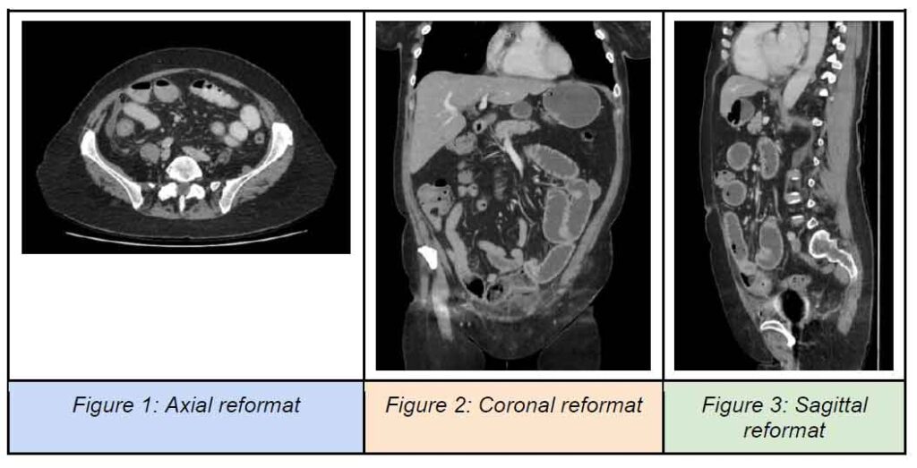
- Multiplanar images generated by thin section scanning (see Figure 1-3 below)
WHAT TO EXPECT DURING A CT ENTEROGRAPHY?
For this procedure, proper bowel preparation is vital. At our practice, patients must fast from all food and drink for 6 hours prior to the exam (5). Patients are then required to drink approximately 1-1.5L of a neutral or low-density oral contrast over 60-90 minutes to achieve adequate luminal distension of the small bowel (5).
This distension is necessary for better assessment of mucosal enhancement, mural thickness, as well as mesenteric vasculature, which is particularly important in the evaluation of Crohn’s disease (5). It is also required as collapsed bowel loops may mimic pathology. During the CT scan, intravenous contrast is administered, and the scan is obtained during the enteric phase where there is peak mucosal enhancement. This technique aims to optimise bowel wall enhancement and contrast resolution between the mucosa and lumen, thereby improving the detection of small bowel abnormalities and pathology (7).
WHAT IS THE ROLE OF CT ENTEROGRAPHY IN MANAGEMENT OF CROHN’S DISEASE?
Diagnosis and assessment of disease extent
CT enterography (CTE) is a highly accurate method for diagnosing and evaluating Crohn’s disease in adults (6). It is particularly useful to assess the severity, extent and location of the disease in the small intestine, which can be challenging with other imaging modalities. This scan can also evaluate any extraluminal manifestations and complications such as strictures, fistulas and abscesses, which is crucial for guiding timely and effective treatment (6).
Treatment decision making
Before commencing medical
treatment or preoperative surgical planning, it is important to use CTE to confirm
the presence of the disease, determine its severity and evaluate any
complications (2). The detailed imaging provided by CTE is valuable by helping
distinguish between active and fibrotic
bowel strictures in patients with Crohn’s
disease, thereby guiding
appropriate treatment decisions which may include medical therapy, surgical resection or endoscopic interventions (3).
Monitoring disease progression and response to treatment
Due to the chronic and relapsing nature of Crohn’s disease, follow up CTE scans are utilised to monitor the disease progression and response to therapeutic strategies (2). By assessing treatment efficacy, appropriate adjustments can be made as needed to the treatment plan (7).
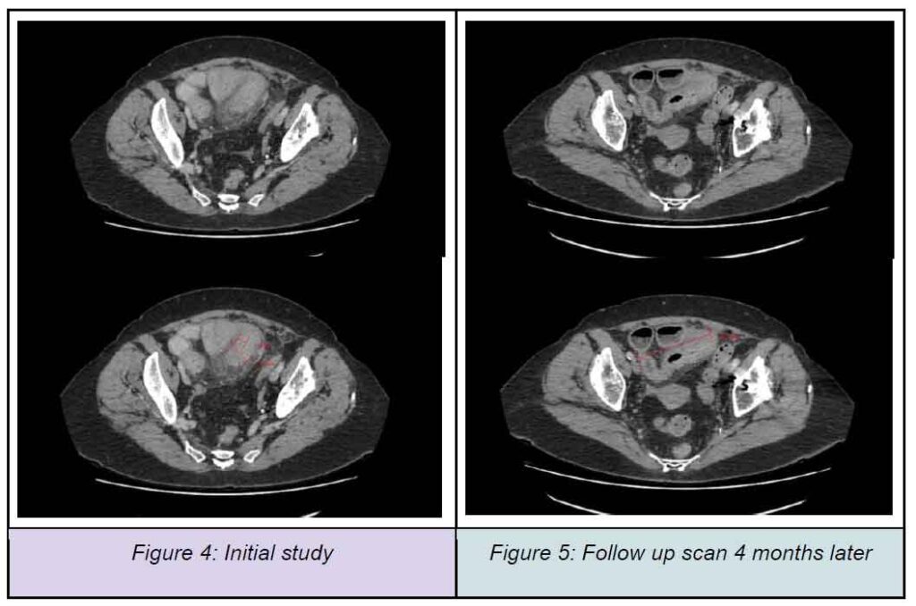
CASE STUDY
A 71-year-old female patient presented to our clinic with a history of Crohn’s disease and increased right sided pain. A CT enterography revealed thickening of the small bowel wall in the ileum, extending over 20 cm to the ileo-caecal junction (see Figure 4). There was also fat stranding in the surrounding area, suggesting possible infectious or an inflammatory bowel condition. Specialist opinion was recommended.
Follow Up Scan
The referring doctor then requested a reassessment of activity following increased therapy. A follow-up CT enterography was performed with multislice sections acquired following oral and intravenous contrast (see Figure 5). Compared to the previous CT study performed four months prior, the results show significant improvements, with less fat stranding and less thickening of the ileum although some inflammatory changes persist.
CONCLUSION
Overall, CTE is a valuable
tool in the comprehensive management of Crohn’s disease,
aiding in diagnosis, monitoring, and treatment planning.
At NIA Diagnostic Imaging, our highly trained staff members and on-site radiologists aim to ensure that all patients have a seamless experience. Please contact us for any further information:
- NIA Glenquarie Radiology
- (02) 9158 8660
- NIA Ingleburn Radiology
- (02) 8104 0803
REFERENCES
- Ali, R. M. M., & Ghonimy, M. B. I. (2021). Diagnostic role of computed tomography enterography (CTE) in assessment of intra-mural and extra-intestinal CT findings in active Crohn’s disease (CD). Egyptian Journal of Radiology and Nuclear Medicine, 52(124). https://doi.org/10.1186/s43055-021-00506-0
- Guglielmo, F. F., Anupindi, S. A., Fletcher, J. G., Al-Hawary, M. M., Dillman, J. R., Grand, D. J., Bruining, D. H., Chatterji, M., Darge, K., Fidler, J. L., Gandhi, N. S., Gee, M. S., Grajo, J. R., Huang, C., Jaffe, T. A., Park, S. H., Rimola, J., Soto, J. A., Taouli, B., Taylor, S. A., … Baker, M. E. (2020). Small bowel Crohn disease at CT and MR enterography: Imaging atlas and glossary of terms. Radiographics, 40(2), 354–375. https://doi.org/10.1148/rg.2020190091
- Ismail, M. S., & Charabaty, A. (2022). Management of Crohn’s stricture: medical, endoscopic and surgical therapies. Frontline Gastroenterology, 13. 524–530.
- Ranasinghe, I. R., Tian, C., & Hsu, R. (2024). Crohn disease. StatPearls Publishing. https://www.ncbi.nlm.nih.gov/books/NBK436021
- Ibrahim, D., Ranchod, A., & Murphy, A. (2023). CT enterography (protocol). Radiopaedia.org. Retrieved June 4, 2024, from https://doi.org/10.53347/rID-30358
- Saade, C., Nasr, L., Sharara, A., Barada, K., Soweid, A., Murad, F., Tawil, A., Ghieh, D., Asmar, K., Tamim, H., & Khoury, N. J. (2019). Crohn’s disease: A retrospective analysis between computed tomography enterography, colonoscopy, and histopathology. Radiography (London), 25(4), 349–358. https://doi.org/10.1016/j.radi.2019.04.007
- Park, M. J., & Lim, J. S. (2013). Computed tomography enterography for evaluation of inflammatory bowel disease. Clinical Endoscopy, 46(4), 327–366. https://doi.org/10.5946/ce.2013.46.4.327