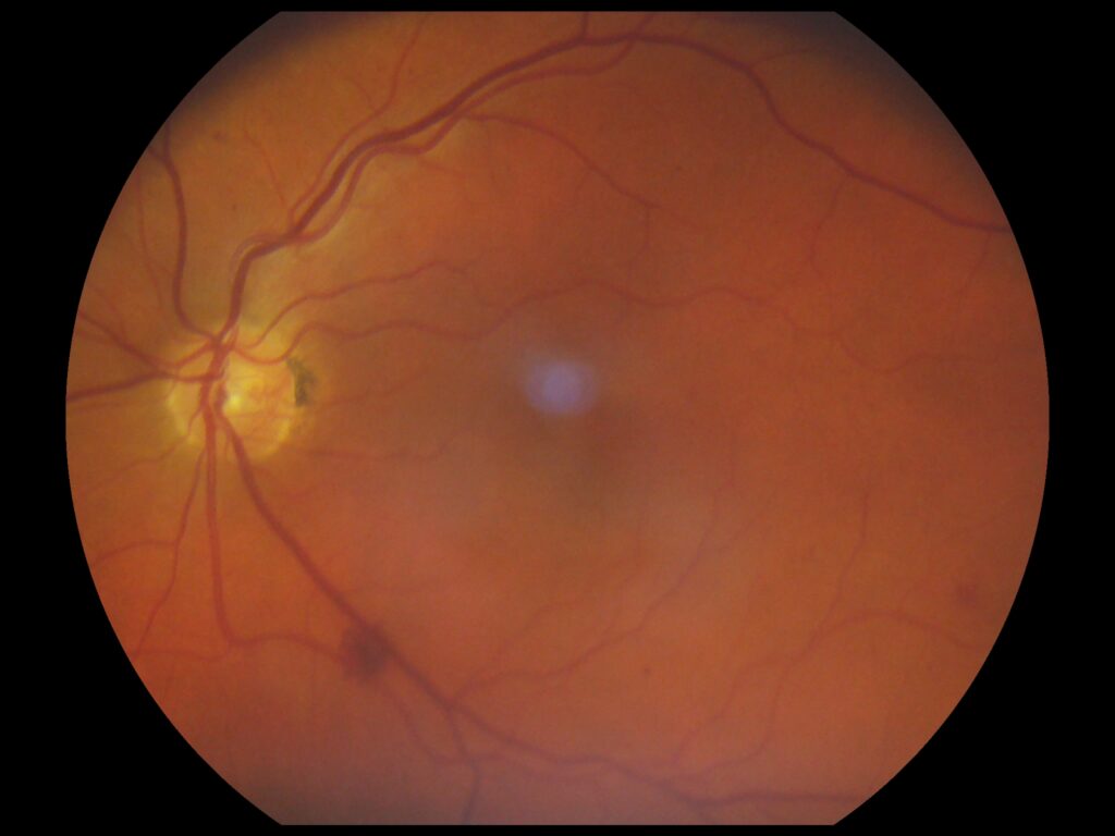
28th September 2024, A/Prof Chee L Khoo

Diabetic retinopathy (DR) is the leading cause of new cases of blindness in patients with diabetes mellitus. In 2020, more than 103 million individuals with diabetes mellitus worldwide were affected by diabetic retinopathy and it is expected that this number will increase to 160 million by 2045 (1). We are all familiar with the association between poor glycaemic control and diabetic retinopathy development and progression. But there is a new angle in the diagnosis and management of diabetic retinopathy. This was presented by Prof Rafael Simo at the most recent EASD meeting in Madrid.
Management of patients with diabetic retinopathy depends on the severity of the retinopathy and whether DME is present. The management when DME is present is messy. It involves photocoagulation, anti-VEGF injections or intravitreal steroid injections. The gist of the management is all about plugging the vascular leak which is thought to be the main cause of damage in diabetic retinopathy and DME. There is increasing body of evidence emerging that early retinal neurodegeneration may precede vascular pathology, suggesting that this neuronal damage may contribute to disease pathogenesis and represent an independent target for intervention. Diabetic retinal neurodegeneration may represent a “preclinical” manifestation of diabetic retinal disease.
The current diagnostic criteria for DR were developed at a time when clinical examination, colour fundus photographs and fluorescein angiography were the mainstays of retinal evaluation. With the advent of ocular coherence tomography (OCT) which was designed to visualise the layers of the macula in cross-section for the diagnosis of macular oedema, alterations in the retinal structure has been detected prior to the onset of DR. OCT has demonstrated thinning of the inner retina (not just limited to the macula ) including the retinal nerve fibre layer, ganglion cell layer, and more variably the inner plexiform layer in patients with type 1 or type 2 DM without DR [2,3].
In addition to thinning of the retinal layers, abnormalities (e.g. functional deficits contrast sensitivity, perimetry testing, multifocal electroretinogram and dark adaptation) have been detected before vascular abnormalities. This neurodegeneration component may be occurring independent of microvascular changes or may actually be contribute to the subsequent microvascular changes.
Mechanisms of Diabetic Retinal Neurodegeneration
In addition to the usual hyperglycaemia induced oxidative stress, inflammation and advanced glycated end products (AGE), there is evidence that glutamate excitoxicity may induced post-synaptic neuronal death (4). Elevated glutamate levels have been found in the vitreous of patients with proliferative diabetic retinopathy [5]. Increased retinal glutamate levels are likely due to reduced expression of the glutamate transporter and resulting impaired glutamate uptake by Muller cells as well reduced glutamate conversion to glutamine by the Muller glia.
Neurons and glia intimately cooperate for energy metabolism in the central nervous system. As neurons invest their ATP in the neurotransmission, glial cells work to support neurons, providing metabolic substrates. Astrocytes, in particular, contribute largely to energy metabolism in CNS. Inflammation as described above, leads to overactivation of glial cells and glial dysfunction (gliopathy), producing first neuroinflammation, maladaptive plasticity, neuronal failure, and later neurodegeneration. Excessive fatty acids from dyslipidaemia causes accumulation in astrocytes which progressively lead to oxidative stress (due to mitochondrial failure), cellular/synaptic dysfunction, neuroinflammation, and, ultimately, cell death.
Similarly, altered glucose transports and metabolism as seen in T2D and insulin resistance, together with dysregulation of neuronal/astrocytic GLUTs and brain insulin resistance, occur in physiological aging, neuroinflammatory and neurodegenerative disorders (6).
Dyslipidaemia, hyperglycaemia and other inflammatory conditions not only cause glial cells activation but they also cause the loss of neuroprotective factors. VEGF and erythropoietin are upregulated while protective neural factors like somatostatin (7), GLP1 and interstitial retinal binding protein (IRBP) are downregulated.
Glial overactivation not only causes neuronal dysfunction and ultimately, death, it is also responsible for microvascular leak. The advent of OCT angiography (OCT-A) technology has allowed greater resolution of the retinal vasculature far beyond what has been detectable by clinical examination or fluorescein angiography. Thus, there is now debate as to whether vascular or neuronal changes occur first. The American Diabetes Association in 2017 described DR is a “highly specific neurovascular complication” of both T1D and T2D (8).
New strategies in management of early DR based on neuroprotection
Current management of DR is solely concentrated on fixing the vascular leak. Apart from addressing the root causes which will reduce glial overactivation and prevent vascular or neuronal damage, what else can we do to prevent the neurodegeneration?
The EUROCONDOR trial examined whether topical administration of two neuroprotective drugs (brimonidine and somatostatin) in patients with early diabetes could prevent or arrest retinal neurodysfunction in patients with type 2 diabetes (9). The primary outcome was the change in implicit time (IT) assessed by multifocal electroretinography between baseline and at the end of follow-up (96 weeks). There was no overall neuroprotective effect of brimonidine or somatostatin but in the subset of patients with preexisting retinal neurodysfunction, IT worsened in the placebo group but remained unchanged in the brimonidine and somatostatin groups. In other words, neuroprotective agents might be beneficial in patients with demonstrated neurodysfunction. What about other neural protective factors that are downregulated?
We are used to using GLP1-RA for management of T2D and weight but GLP1-RA exerts neuroprotective effects in both the central and peripheral nervous system. In genetically modified diabetic mice, GLP1 administered topically (i.e. eye drops) have been shown to upregulate glutamate aspartate transporter (GLAST) which is responsible for clearing glutamate from the extracellular space which in turn prevent neuronal dysfunction. In addition, GLP1-RA topically was shown to have microvascular protection (10). There were no effects on plasma glucose from topical GLP1-RA. Obviously, the problems with GLP1-RA, like in plasma is the rapid degradation by DPP4 inhibitors.
Topical sitagliptin has been shown to do achieve the same as topical GLP1-RA (11). In addition , sitagliptin has been shown to prevent the downregulation of protective crucial presynaptic proteins (12).
In summary, when we think of diabetic retinopathy, we think about microvascular leakage and all disease staging is based on the degree of the leak, the ischaemia leading to neovascularisation and whether the macula is involved. Treatment up until now is about stemming the microvascular leak. But research over the last decade has uncovered neurodegeneration and microvascular changes can predate the vascular leak. Various topical eye drops (including DPP4 inhibitors) look promising, at least in animal studies, in preventing neurodegeneration and microvascular damage before early diabetic retinopathy is even diagnosed. We await human trials.
References:
- Teo ZL, Tham YC, Yu M, et al. Global prevalence of diabetic retinopathy and projection of burden through 2045: systematic review and meta-analysis. Ophthalmology 2021; 128(11):1580–1591
- Carpineto P, Toto L, Aloia R, et al. Neuroretinal alterations in the early stages of diabetic retinopathy in patients with type 2 diabetes mellitus. Eye (Lond). 2016;30(5):673–679.
- van Dijk HW, Verbraak FD, Kok PH, et al. Decreased retinal ganglion cell layer thickness in patients with type 1 diabetes. Invest Ophthalmol Vis Sci. 2010;51(7):3660–3665
- Mizutani M, Gerhardinger C, Lorenzi M. Muller cell changes in human diabetic retinopathy. Diabetes. 1998;47(3):445–449 7. Magistretti PJ, Allaman I. Lactate in the brain: From metabolic end-product to signalling molecule. Nat Rev Neurosci. 2018;19:235–249
- Ambati J, Chalam KV, Chawla DK, et al. Elevated gamma-aminobutyric acid, glutamate, and vascular endothelial growth factor levels in the vitreous of patients with proliferative diabetic retinopathy. Arch Ophthalmol. 1997;115(9):1161–1166
- Scherer T, Sakamoto K, Buettner C. Brain insulin signalling in metabolic homeostasis and disease. Nat Rev Endocrinol. 2021;17:468–483
- Carrasco E, Hernández C, Miralles A, et al. Lower Somatostatin Expression Is an Early Event in Diabetic Retinopathy and Is Associated With Retinal Neurodegeneration. Diabetes Care 2007;30(11):2902–2908
- Solomon S, Chew E, Duh E, et al. Diabetic Retinopathy – A Position Statement by the American Diabetes Association. Diab Care 2017, 40, 412-418.
- Simó R, Hernández C, Porta M, et a. European Consortium for the Early Treatment of Diabetic Retinopathy (EUROCONDOR). Effects of Topically Administered Neuroprotective Drugs in Early Stages of Diabetic Retinopathy: Results of the EUROCONDOR Clinical Trial. Diabetes. 2019 Feb;68(2):457-463.
- Hernandez C, Bogdanov P, Corraliza L. et al. Topical administration of GLP1-RA Prevents Retinal Neurodegeneration in experimental Diabetes. Diabetes 2016, 65: 172-187
- Hernandez C, Bogdanov P, Sola-Adell C, et al. Topical administration of DPP4 inhibitors Prevents Retinal Neurodegeneration in experimental Diabetes. Diabetologia 2017, 60:2285-2298
- Ramos H, Bogdanov P, Sabater D, et al. Neuromodulation Induced by Sitagliptin: A New Strategy for Treating Diabetic Retinopathy Biomedicines 2021, 9 (12): 2172
