
14th November 2025, NIA Diagnostic Imaging
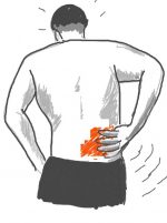
Lower back pain (LBP) remains one of the most common presentations in general practice and a major cause of disability worldwide. (3) While most cases are non-specific and self-limiting, imaging can be valuable when serious underlying pathology is suspected or symptoms persist beyond the usual recovery period. The two key imaging modalities utilised to diagnose LBP are computed tomography (CT) and magnetic resonance imaging (MRI). Understanding the benefits and limitations of these modalities is crucial for making appropriate, evidence-based decisions that result in an accurate diagnosis as well as patient care and satisfaction. (1)
CT VS MRI: BRIEF OVERVIEW
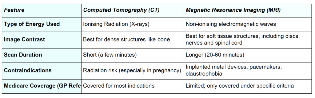
Although both CT and MRI generate comprehensive cross-sectional pictures, each modality possesses distinct benefits and limitations in the clinical assessment of lower back pain. The following table summarises the various features offered by each modality:
CHOOSING THE RIGHT MODALITY
In diagnosing back pain, the choice between MRI and CT is dependent on the suspected cause of pain. Ultimately, MRI is the golden standard for assessing soft tissue structures, including vertebral discs, nerves, and muscles. Some conditions involving these structures would include herniated discs, spinal stenosis and degenerative disc disease. On the other hand, CT is more suited to examining and detecting bone-related issues, including fractures or spinal malalignment. Due to its speed and high resolution for bony structures, CT is often the preferred option in emergency settings, especially for trauma cases. (2,5)
CASE STUDY
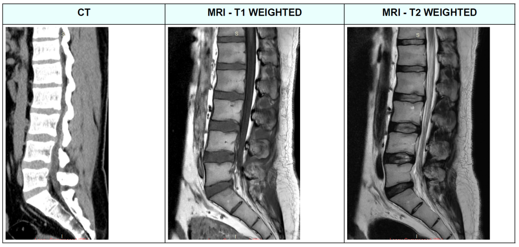
A 38-year-old male presented to our clinic with bilateral leg radiculopathy and lower back pain after being assaulted at work. An initial CT scan revealed that the patient had multilevel disc bulging, with no evidence of fractures or nerve root impingement.
A week later, the patient returned to our clinics and underwent an MRI scan. The MRI demonstrated that the patient
had a disc desiccation and an annular disc tear at L5/S1, as well as disc bulging at L3/4, potentially impinging on the
origin of the right L4 nerve root.
CONCLUSION
In conclusion, the decision to use CT or MRI for diagnosing lower back pain should be guided by the clinical context. While CT is particularly useful for identifying bone-related issues, MRI is better suited for assessing soft tissue
structures, including discs, nerves, and the spinal cord. As demonstrated in the case study, MRI is capable of depicting soft tissue details that were not apparent on the CT scan. Therefore, choosing the right imaging technique is crucial for achieving an accurate diagnosis, which in turn ensures effective management of the patient’s condition.

- REFERENCES
- Australian Commission on Safety and Quality in Health Care. (2022). Low Back Pain Clinical Care Standard. https://www.safetyandquality.gov.au/sites/default/files/2022-08/low_back_pain_clinical_care_standard.pdf
- Hussain, O., Kaushal, M., Agarwal, N., Kurpad, S., & Shabani, S. (2023). The Role of Magnetic Resonance Imaging and Computed Tomography in Spinal Cord Injury. Life, 13(8), 1680. https://doi.org/10.3390/life13081680
- Hutchins, T. A., Peckham, M., Shah, L. M., Parsons, M. S., Agarwal, V., Boulter, D. J., Burns, J., Cassidy, R. C., Davis, M. A., Holly, L. T., Hunt, C. H., Khan, M. A., Moritani, T., Ortiz, A. O., O’Toole, J. E., Powers, W. J., Promes, S. B., Reitman, C., Shah, V. N., & Singh, S. (2021). ACR Appropriateness Criteria® Low Back Pain: 2021 Update. Journal of the American College of Radiology, 18(11), S361–S379. https://doi.org/10.1016/j.jacr.2021.08.002
- RACGP – Imaging in adults with acute low back pain. (2018). Racgp.org.au. https://www.racgp.org.au/clinical-resources/clinicalguidelines/key-racgp-guidelines/view-all-racgp-guidelines/first-do-no-harm/gp-resources/imaging-in-adults-with-acute-low-backpain
- Ruiz Santiago, F., Láinez Ramos-Bossini, A. J., Wáng, Y. X. J., Martínez Barbero, J. P., Espinosa, J. G., & Martínez Martínez, A. (2022). The value of magnetic resonance imaging and computed tomography in the study of spinal disorders. Quantitative Imaging in Medicine and Surgery, 12(7), 3947–3986. https://doi.org/10.21037/qims-2022-04
- Stallard, J. (2019, May 10). CT vs MRI: What’s the Difference? And How Do Doctors Choose Which Imaging Method to Use? Memorial Sloan Kettering Cancer Center. Www.mskcc.org. https://www.mskcc.org/news/ct-vs-mri-what-s-difference-and-how-dodoctors-choose-which-imaging-method-use