
!2th July 2026, NIA Diagnostic Imaging
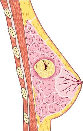
Breast fibroadenomas are common benign breast tumours that primarily occur in women of reproductive age (1). They are composed of both glandular and connective tissue tissue, and typically present as a well-defined, firm and mobile lump (4). Though fibroadenomas are not cancerous, they are clinically significant due to the potential to mimic the clinical and imaging features of breast cancer. Therefore, it is essential to establish the correct diagnosis to guide appropriate management and ensure timely care.
What causes breast fibroadenomas?
The cause of fibroadenomas is not fully understood, however, it appears that fibroadenomas are hormone-related with increased growth of glandular and connective tissue. For this reason, fibroadenomas may proliferate during pregnancy or other periods of hormonal change, and become smaller after menopause (1).
What are the signs and symptoms?
Many fibroadenomas may be discovered incidentally during routine breast imaging. Common signs and symptoms include: (1, 3)
- Painless, firm breast lump
- May be mobile underneath the skin
- Changes in appearance or contour of breast
What is the first diagnostic tool?
Diagnosis of fibroadenomas is based on a combination of clinical examination, breast imaging and histopathological confirmation, where necessary (3). Breast ultrasound is the preferred first-line imaging tool for young women with dense breast tissue due to its widespread availability, does not use ionising radiation and real time assessment (4). Mammography may be used as adjunct, particularly in older women or when additional imaging is indicated.
Ultrasound helps clinicians (3):
- Assessment of size, location and internal characteristics of the lesion
- Monitor changes with serial ultrasound examinations
- Guide biopsy procedures when tissue sampling is required
- On ultrasound, fibroadenomas typically appear as (1, 3):
- Well-defined, oval or round masses
- Uniformly hypoechoic
- Smooth, circumscribed margins
What are the clinical management and treatment options?
Clinical management and treatment planning of fibroadenomas are individualised, and depend on the size, patient symptoms, age, imaging findings and personal preference.
- Observation: Most fibroadenomas do not require surgical treatment and can be managed conservatively with regular clinical assessment and follow up imaging to monitor for changes.
- Biopsy: For uncertain imaging findings, increasing size or atypical features, a biopsy (such as fine-needle aspiration or core needle biopsy) may be performed for definitive diagnosis.
- Surgical and minimally invasive treatment: For fibroadenomas that require surgical management, various options include excisional biopsy, lumpectomy and cryoablation in selected patients.
Conclusion
At NIA Diagnostic Imaging, high-resolution breast ultrasound using GE LOGIQ™ E10 ultrasound systems provides detailed assessment of breast tissue, helping identify and characterise fibroadenomas while supporting accurate diagnosis and appropriate patient management.

References:
- Ajmal, M., Khan, M., & Van Fossen, K. Breast fibroadenoma. StatPearls Publishing. https://www.ncbi.nlm.nih.gov/books/NBK535345/
- Nnadi, C. (2025). Fibroadenoma [Image]. Med Bullets. https://step2.medbullets.com/gynecology/120421/fibroadenoma
- Pandit, P., Murkey, S. P., Agarwal, A., Jaiswal, A., & Agrawal, S. (2023). Understanding fibroadenoma of the breast: A comprehensive review of pre-operative and post operative clinicopathological correlations. Cureus, 15(12). https://doi.org/10.7759/cureus.51329
- Ramala, S. R., Jr, Chandak, S., Chandak, M. S., & Annareddy, S. (2023). A comprehensive review of breast fibroadenoma: Correlating clinical and pathological findings. Cureus, 15(12). https://doi.org/10.7759/cureus.4994