
9th September 2026, NIA Diagnostic Imaging
Acute knee injuries commonly occur as a result of direct blows to the joint, such as motor vehicle accidents, but can also occur after sudden twisting, instability events, or falls (4). After the injury, patients often clinically present with any or all of the following: focal or generalised knee pain, locking of the knee, joint effusion or inability to bear weight. There are multiple soft tissue or bony structures in the knee that can be injured. The severity of the injury can range from mild sprains or localised bruising to significant soft tissue (including ligament or cartilage tears) and fractures. Choosing the right imaging modality can be tricky.
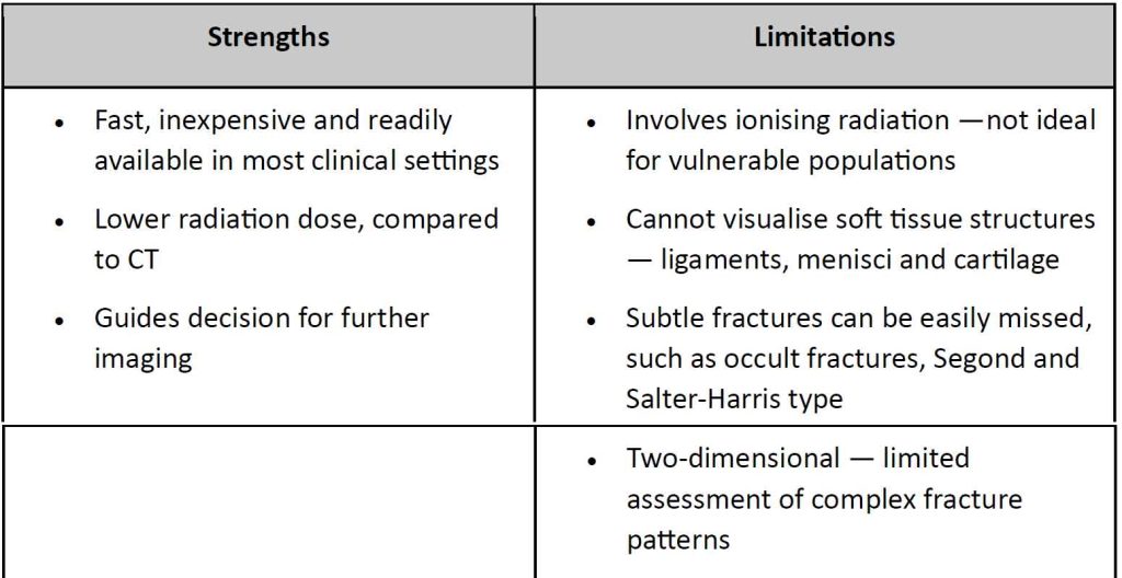
Plain X-Ray
X-ray is the standard first-line imaging modality for acute knee injuries. Capable of detecting:
- Fractures – patella, tibial plateau, femoral condyle
- Avulsion fractures
- Loose bodies within the joint
- Bony malalignment
- Joint effusion – indicative of further underlying pathology
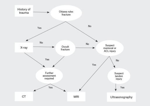
The Ottawa Knee Rules help GPs identify which patients genuinely need X-ray, reducing unnecessary radiation exposure. (3)
X-ray is recommended if the patient has any of the following:
- Age ≥55 years
- Isolated patella tenderness
- Fibula head tenderness
- Inability to flex knee to 90 degrees
- Inability to bear weight immediately after injury and inability to take four steps in the emergency department
If none of these criteria are met, imaging can often be safely deferred, and conservative management with follow-up review may be appropriate. If symptoms persist or worsen, imaging can be arranged at that point.
Magnetic Resonance Imaging (MRI)
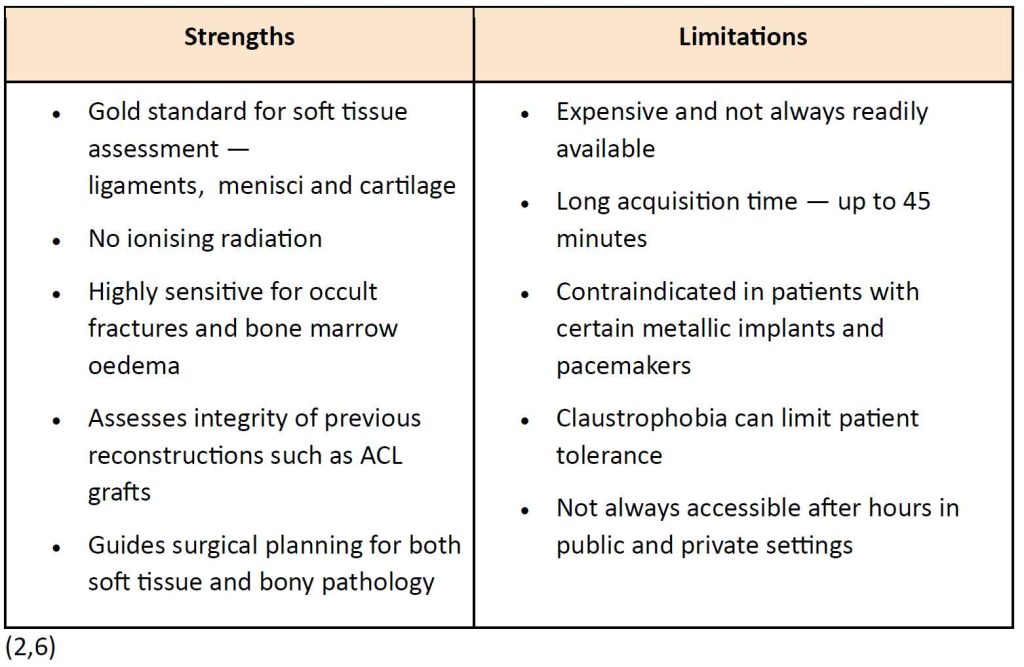
Magnetic resonance imaging (MRI) is the gold standard for evaluating and diagnosing soft tissue injuries in the knee. (6)
Key roles of MRI in acute knee injuries include:
- Identification of ACL, PCL, MCL and LCL tears
- Detection of meniscal tears — medial and lateral
- Assessment of articular cartilage damage and osteochondral lesions
- Identification of bone marrow oedema and bone bruising
- Detection of occult fractures not visible on x-ray
MRI also plays an important role in surgical planning and assisting clinicians by:
- Guiding surgical decision-making for both soft tissue and bony pathology
- Evaluating the integrity of previous reconstructions such as ACL grafts
- Providing comprehensive assessment of all structures before arthroscopy
- Determining whether surgical or conservative management is most appropriate
Computed Tomography (CT)
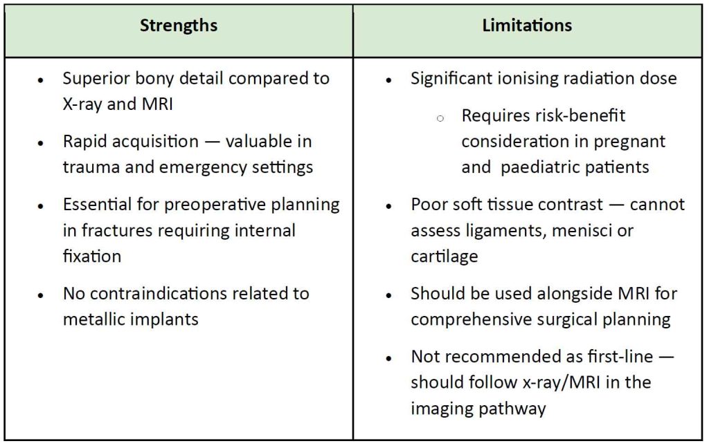
Computed Tomography (CT) is not recommended as a first-line imaging modality for most acute knee injuries.
Instead, CT serves a complementary role, sitting between plain radiography and MRI in the imaging pathway. (5)
Key indications include:
- Evaluation of occult fractures not visible on initial X-ray
- Characterisation of known fractures, particularly for preoperative planning
- 3D reconstruction for complex fracture patterns
- Assessment of articular depression, fragment orientation and comminution.
Ultrasound
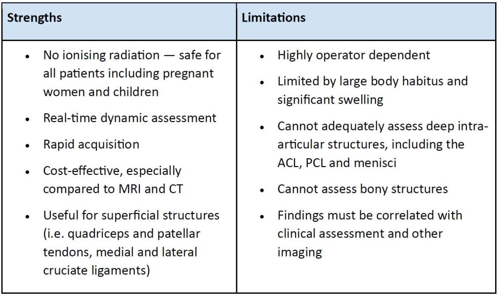
Despite MRI remaining the gold standard, ultrasound is gaining popularity due to its real-time imaging, accessibility, time-efficiency and safety. (5)
Key indications include:
- Pathology within superficial structures — patellar and quadriceps tendons and collateral ligaments
- Detection of joint effusion and haemarthrosis post-trauma; indicative of further underlying pathology
In the private setting, ultrasound remains a practical alternative for soft tissue imaging, where MRI availability and costs are barriers.
As per the Royal Australian College of General Practitioners, Clinical Imaging selection in acute knee injuries is guided by the findings of physical examination and clinical diagnosis. (5)
- X-ray: first-line for trauma when indicated by clinical decision tools
- MRI: gold standard for soft tissue structures and internal derangement
- CT: complex fracture characterisation and surgical planning
- Ultrasound: superficial soft tissue injuries or when MRI is inaccessible/contraindicated

At NIA Diagnostic Imaging, we offer all imaging modalities across three conveniently located sites. We accept urgent appointments to support timely assessment of acute knee injuries.
REFERENCES
- Australian Primary Health Care Nurses Association. (2017). Acute ankle and knee injuries: Limited role for imaging. Primary Times, 17(1).
- Choudhary, N., Nagar, A., Singh, S., & Lakhar Guru, B. (2025). Role of MRI in diagnosis of knee injuries: An observational study. International Journal of Medicine and Public Health, 15(1), 43–48.
- Haque, T., & Yao, K. (2012). The Ottawa knee rules: A useful clinical decision tool. Australian Family Physician, 41(4), 229–231. https://www.racgp.org.au/afp/2012/april/the-ottawa-knee-rules
- Queensland Health. (2024, February 13). Knee injury (acute). Queensland Government. https://www.health.qld.gov.au/cpc/orthopaedics/acute-knee-pain
- Ridley, U., & Ridley, L. (2020). Imaging of the knee: Common acute presentations to general practice. Australian Journal of General Practice, 49(6). https://doi.org/10.31128/AJGP-10-19-5120
- Shetty, N. D., Bora, N., & Shelar, S. S. (2024). The role of magnetic resonance imaging in the evaluation of knee pain. Cureus, 16.