
12th November 2026, NIA Diagnostic Imaging
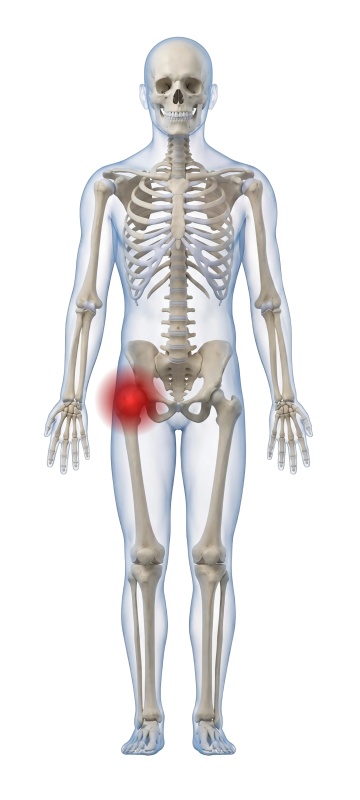
Hip pain can have multiple causes, including intra-abdominal, intra-articular (degenerative changes and inflammation), extra-articular (muscle injury and bursitis). Hip pain can usually be localised in one of the three regions: anterior and groin, lateral, or posterior and buttock. As there are numerous causes of hip pain, patient history, localisation of pain and physical examination are critical for deciding on the approach to pain management. Imaging can be further utilised to confirm and rule the causes of hip pain, as well as precisely administer corticosteroid treatment.
Osteoarthritis and CT-Guided Cortisone Injections
In adults over 45 years, osteoarthritis (OA) is a common cause of hip pain, and one in four adults will develop symptomatic osteoarthritis in their lifetime. OA can have a significant impact on patient life through decreasing levels of physical activity, and it usually manifests anteriorly or as buttock pain. Plain radiography is helpful in diagnosing osteoarthritis by assessing the narrowing of joining spaces, particularly the articulation between the femoral head and the acetabulum, sclerosis present in the joint space and presence of osteophytes.
Intraarticular corticosteroid (IACS) injections are available to elevate hip pain caused by osteoarthritis through immunosuppressive and anti-inflammatory mechanisms of action. The injections are performed under CT – the needle is precisely guided into the intraarticular space, and the corticosteroid medication is administered directly into the joint capsule.
Randomised control trials presented in literature reviews indicate that injections can provide substantial pain relief and improvement in mobility for up to 12 weeks. It is particularly applicable to patients for whom Total Hip Arthroplasty is not clinically indicated or those that have contraindication to surgery. Although there is generally no preparation needed, some contraindications should be addressed including diabetes, anticoagulants, infection and allergies. Care must also be taken post injection to protect the area from infection.
Bursitis and Ultrasound-Guided Injections
Bursitis (trochanteric bursitis) is another common cause of hip pain that can cause significant pain and mobility impairment, attributed to the inflammation of a bursa stemming from overuse, infection, trauma or inflammation. Although plain radiography is commonly used to detect trauma or a foreign body that can attribute to swelling and inflammation, ultrasound provides extensive benefit for tissue visualisation and real-time imaging observing tissue changes around the bursa with movement (5).
Bursitis is generally associated with the lateral hip pain and focal tenderness over the greater trochanter, although it can also manifest as buttock or thigh pain. Ultrasound-guided injections are precise and quick interventional procedures. Corticosteroid injections for bursitis have been found to elevate pain for several weeks to months. These injections are indicated for patients who fail conservative management, which include NSAID’s, exercise, physical therapy and weight reduction.
Contraindications to corticosteroid injections for bursitis include systemic infection, cellulitis over the injection site, and significant immunocompromise. Anticoagulation medications may be continued.
At NIA Diagnostic Imaging, pain management injections are Bulk-Billed under Medicare. This ensures all patients have affordable access to pain relief, thus improving their quality of life.
References
- Chamberlain R. Hip Pain in Adults: Evaluation and differential diagnosis. American Family Physician [Internet]. 2021 [Cited 2026 Nov 4]; 103(2):81-89. Available from: aafp.org/afp/2021/0115/p81#afp20210115p081-b4
- Choueiri M, Chevalier X, Eymard F. Intraarticular Corticosteroids for Hip Osteoarthritis: A Review. Cartilage [Internet]. 2020 [Cited 2026 Nov 5]; 13(1 Suppl):122S-131S. Doi: 10.1177/1947603520951634.
- Santiago FR, Chinchilla AS, Ansari A, Alvarez LG, Garcia MMC, Martinez AM, Sanchez JT. Imaging of Hip Pain: From Radiography to Cross-Sectional Imaging Techniques. Radiology Research and Practice [Internet]. 2016 [Cited 2026 Nov 7]; 2016:6369237. Doi: 10.1155/2016/6369237
- Williams CH, Jamal Z, Stenard BT. Bursitis. Treasure Island (FL): StatPearls Publishing [Internet]. 2023 [Cited 2026 Nov 7]; Available from: https://www.ncbi.nlm.nih.gov/books/NBK513340/