28th June 2020, Dr Chee L Khoo

In type 1 diabetes (T1D), the β-cell die rapidly from a massive immunological assault and practically all the β-cells are quickly lost and hence, there is an absolute deficiency of insulin secretion. Using auto-antibody screening, we can define T1D. Do you realise that we don’t actually have a definition for type 2 diabetes (T2D). If we exclude T1D, monogenetic diabetes and secondary diabetes, we call the rest T2D. Over the past decade, major advances have been made in the studies of islet morphology and human β-cell gene expression in T1D and T2D and the endoplasmic reticulum stress signalling that contributes to β-cell failure in T1D and T2D. A dead β-cell is a dead β-cell but understanding the mechanisms behind β-cell failure is critical to prevent or revert disease both in T1D and T2D.
Most of our knowledge on islet morphology and the mechanisms leading to diabetes mellitus were based on limited studies of human pancreata collected at necropsy and on animal models such as non-obese diabetic (NOD) mice, ob/ob and db/db mouse models of obesity and T2D, and the GK rat model of T2D, which reproduce the human disease to a rather limited extent.
The development of the Network for Pancreatic Donors with Diabetes as well as the systematic organisation and expansion of the Exeter Archival Diabetes Biobank have already provided access to ~500 well- preserved pancreata from individuals with T1D and non- diabetic controls from both necropsy and organ donors, with different ages of disease onset and duration.
Eizirik, Pasquali and Cnop (1) recently reviewed findings over the last 15 years on studies performed on human beta cells in both T1D and T2D.
Why do the β-cell die?
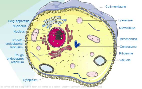
Endoplasmic reticular stress (ER stress) develops when the demand to synthesise and process proteins in the ER exceeds capacity. This situation can arise as a result of a variety of perturbations such as, for example, expression of a mutant protein, translational misreading, increased demand for protein synthesis, ATP shortage, ER Ca2+ depletion, impaired N- linked glycosylation, alterations in the oxidising environment of the ER, shortage of ER folding enzymes or chaperones and accumulation of cholesterol or saturated lipids in the ER membrane.
Evidence has accumulated that ER stress contributes to β-cell failure in T1D and T2D. However, the signalling that initiates the damage differs, with predominantly IRE1- mediated β-cell damage in T1D and PERK–eIF2α- mediated β- cell damage in T2D. In vitro studies have identified potential triggers of β-cell ER stress in T2D, notably high glucose or saturated FFA levels. The ability to modulate β-cell ER stress could be an interesting adjuvant treatment to new approaches aiming to decrease immune-mediated β-cell death in early T1D.
T1D – the bi-directional immune assault
T1D is caused by autoimmune-mediated β-cell dysfunction and apoptosis. We now know that the auto-immune disease is the consequence of a complex dialogue between invading or resident macrophages and T cells and β-cells . The immune cells release chemokines and cytokines into the islet micro-environment and trigger local inflammation leading to progressive β-cell dysfunction and ultimately cell death via apoptosis. The immune attack is not a one-way street. β-cells generate signals when stressed, injured or dying and these signals attract and activate immune cells adding to the inflammation. In other words, β-cells play an active role in their own demise in T1D!
Not all responses induced by these cytokines are deleterious to β-cells. For example, both IFNα and IFNγ upregulate β-cell expression of PDL1 and HLA-E64, proteins that respectively provide a negative feedback to T cells and natural killer cells and protect β-cells against immune-mediated cell death.
A feasible therapeutic approach might involve preserving protective signals downstream of IFNs (such as PDL1) whilst blocking deleterious signals (such as HLA class I overexpression, ER stress and chemokine production).
The pathogenesis of
T2D
The histology of pancreatic islets in patients with T2D comprises an ~40%
reduced β-cell mass, increased β-cell apoptosis, greater amyloid deposition and
reduced pancreatic insulin content compared with that of non-diabetic
pancreatic islets. β-cell dysfunction though, is a lot more severe (up to 80%
reduction) than the reduction in beta cell mass suggesting the that β-cell
dysfunction is an early player in T2D pathogenesis and occurs independently of
β-cell loss.
The human β-cell mass is established in the first 2–3 decades of life and that, subsequently, β-cells age with the body. Unlike T1D, β-cell apoptosis in T2D is an infrequent event. Although the end result of β-cell dysfunction and β-cell death is similar in both T1D and T2D, their causative factors differ. Cytokines lead to β-cell dysfunction and death in T1D, whereas free fatty acids (FFAs) might elicit ER stress and thereby impair β-cell function and survival in T2D.
Accumulating evidence suggests that accelerated β-cell ageing and senescence occurs in T2D. In keeping with the long lifespan of β-cells, one study showed that β-cell and α-cell telomere shortening is most pronounced before age 20 years and flattens thereafter, pointing to
replicative senescence of post-mitotic adult β-cells. More marked telomere attrition was seen in β- cells in T2D.
Dysfunction before death
Some individuals from families affected by T1D show evidence of β-cell dysfunction, such as decreased first phase glucose-stimulated C-peptide release or increased circulating proinsulin–insulin ratios, in the absence of β-cell autoantibodies. This observation suggests that β- cell dysfunction could actually precede the autoimmune assault in T1D or might reflect ‘scars’ of a previous, resolved autoimmune episode. Thus, is it possible that stressed and dysfunctional β-cell release signals which initiate the immune attack?
At disease onset, β-cell loss is far from complete. In some patients with recent- onset T1DM who died in ketoacidosis, the remaining ß-cell mass approached 40–50% of normal, suggesting that severe immune- mediated ß-cell dysfunction precedes actual ß-cell death. In imaging mass cytometry studies, loss of the β-cell markers insulin, proinsulin and amylin, precedes β-cell death.
Islets obtained from NOD mice affected by severe insulitis are dysfunctional upon isolation, however, they regain function once freed from the infiltrating immune cells after 7 days in culture. β-cells from four donors with T1D (2–7 years of duration) had preserved insulin release in response to glucose, when expressed as insulin secretion per insulin content, but showed defective first- phase insulin release.
Insulin release, as evaluated by stimulated urinary C-peptide, is detectable in 80% of patients with T1D after a mean disease duration of 21 years, most often at very low levels. Even in some C-peptide-negative patients with T1D still produce and release proinsulin, suggesting that surviving insulin- producing cells have defective proinsulin–insulin conversion.
The Genetics of Diabetes
During the last decades, the human genome of thousands of individuals was scanned in search of DNA sequence variants associated to common traits and diseases. Genome wides association studies (GWAS) revealed >400 distinct genetic signals associated with T2D and >50 influencing T1D. However, the most common associated variants with T1D and T2D have only modest effects on disease risk. In the context of T1D pathogenesis, genetic variants could affect the speed of functional β-cell loss following the appearance of auto-antibodies.
T1D has a strong heritable component (twin concordance rate up to 70% and sibling risk of approximately 8%, which enables disease prediction based on individual genetic background. A genetic risk score was developed based on 67 single nucleotide polymorphisms to include HLA DR- DQ haplotype interactions as well as non- HLA single nucleotide polymorphisms. The resulting score allowed discrimination of T1D in the UK Biobank dataset with an outstanding accuracy.
In T2D, the data emerging from GWAS indicate that a substantial fraction of the association signals are driven by dysregulation of β-cell development and insulin secretion, as opposed to influencing the tissues of insulin action such as fat, muscle and liver.
ß-cell failure is the central event in both T1D and T2D but the pathways that lead to this failure are different. ß- cell dysfunction and death in T1DM is mostly immune mediated, whereas metabolic stress has a clear role in the progressive loss of functional ß- cell mass in patients with T2D.
Non-coding genes
The human genome contains large numbers of non-coding genes which govern the function and fate of target cells (including islet cells). GWAS have implicated islet-specific non-coding function with genetic susceptibility for T2D. Attempts have been made to bridge these gaps in knowledge by studies applying state- of- the- art techniques to reconstruct regulatory relationships between distal regulatory elements and their target genes.
The plasticity of islet cells
Dedifferentiation and/or trans-differentiation have also been implicated in the β-cell pathology of T2D. α-cells can differentiate into β-cell and vice versa. The same can occur in δ-cells. Glucagon and insulin co- expressing cells are increased in islets from patients with T2DM (3–4% versus 0.5–3% in non- diabetic islets). The impact of β-cell de-differentiation and trans-differentiation on functional β-cell mass in T2D remains uncertain.
Can we translate the growing understanding of β-cell fate in diabetes mellitus into novel biomarkers that allow us to predict disease or to follow β- cell loss and response to therapy? Can novel approaches be developed to improve ER function in both forms of diabetes mellitus? Can the addition of novel therapies aiming to protect β- cells in T1D improve the limited benefits of ongoing attempts to revert disease based on targeting the immune system only?
We have come a long way in understanding β-cell demise but we have a long way to go yet.
References:
Eizirik, D.L., Pasquali, L. & Cnop, M. Pancreatic β-cells in type 1 and type 2 diabetes mellitus: different pathways to failure. Nat Rev Endocrinol 16, 349–362 (2020). https://doi.org/10.1038/s41574-020-0355-7
