
12th August 2026, NIA Diagnostic Imaging
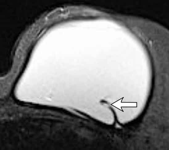
Breast implant rupture develops when silicone or saline implants fail, causing the leakage of their contents. 10% to 15% of implants will rupture within 10 – 15years. The risk increased with implant age, type and surgical factors. At NIA Diagnostic Imaging, we offer Bulk-Billed Breast Ultrasound and Mammography. Breast MRI is also available at NIA Diagnostic Imaging.
CAUSES OF IMPLANT RUPTURE
- Implant age
- Trauma
- Manufacturing defects
- Incompetent valves
- Underfilling of the implant
A fibrous capsule is scarring created by the body around the implant as part of a normal reaction to the foreign implant. There are two types of rupture; intracapsular rupture and extracapsular rupture.
TYPES OF IMPLANT RUPTURE
INTRACAPSULAR RUPTURE
- The implant rupture is contained within the fibrous capsule.
EXTRACAPSULAR RUPTURE
- Extravasation of the implant content outside the fibrous capsule.
- Isotonic saline solution in saline implant rupture can be safely reabsorbed by the body over time.
- Silicone rupture can lead to local tissue reaction and scar formation.
SYMPTOMS OF IMPLANT RUPTURE
In saline implant rupture the most common sign is gradual or acute volume loss and can be easily detected on clinical examination. Silicone implant rupture is harder to recognise on clinical examination. Extracapsular rupture can sometimes cause a palpable mass or irregularity of the breast which is caused by inflammation and formation of granulomatous tissue. Patients may experience vague pain, tenderness or increase firmness in the breast. Therefore, further investigation with imaging is required.
IMAGING
MRI
The gold standard for the investigation of implant rupture with reported sensitivity of 72–94% and specificity of 85–100%. However, it is expensive, time-consuming, uncomfortable for the patient and there are contraindications
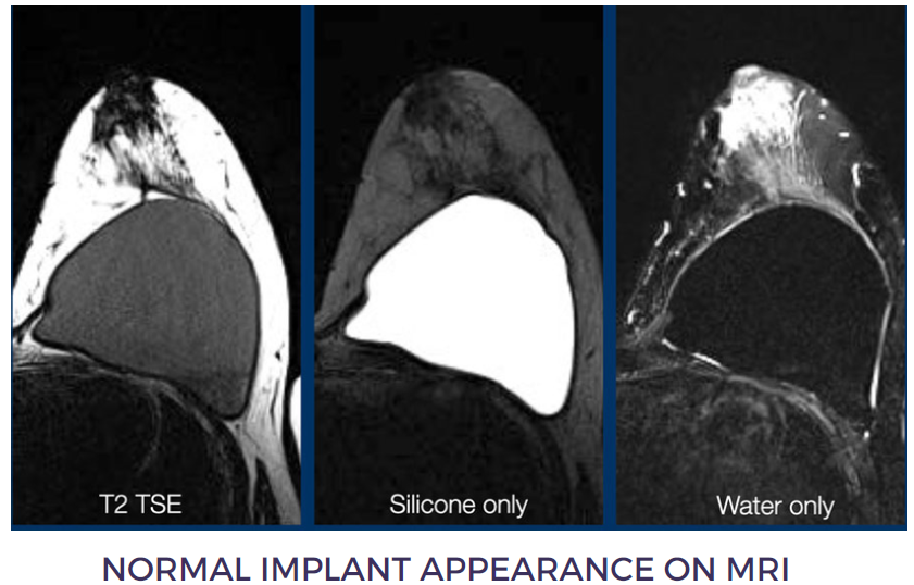
INTRACAPSULAR RUPTURE
Silicone is seen within the implant fold (Image 2).
- Teardrop sign: walls of the fold are touching each other (1)
- Keyhole or Noose sign: walls of the fold are not touching each other (2 & 3)
- Linguine sign: ruptured envelope wall created curvilinear lines that resemble Linguine pasta (Image 3).
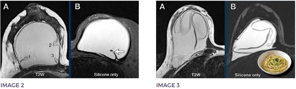
EXTRACAPSULAR RUPTURE
Silicone is seen outside the capsule
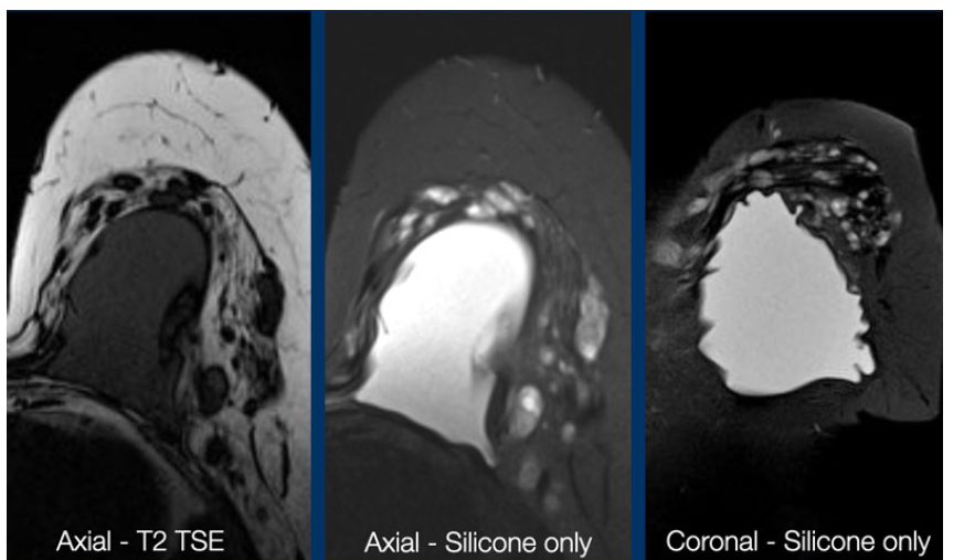
ULTRASOUND
- Popular alternative to MRI in the investigation of implant rupture as it is time and cost efficient.
- However, it is highly operator dependent and has a decrease in sensitivity in the presence of capsular contracture.
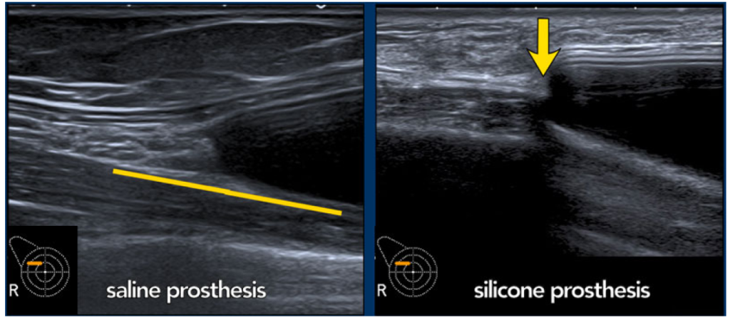
NORMAL IMPLANT
INTRACAPSULAR RUPTURE
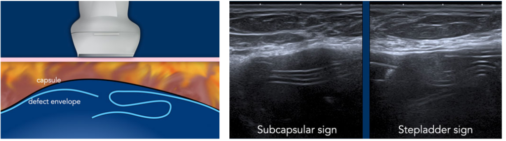
- Subcapsular sign: Parallel lines within the content of the breast implant, not following the outer envelope capsular complex.
- Stepladder sign: multi layer parallel lines inside the content of the implant (similar to Linguine sign on MRI) (Image 7).
EXTRACAPSULAR RUPTURE
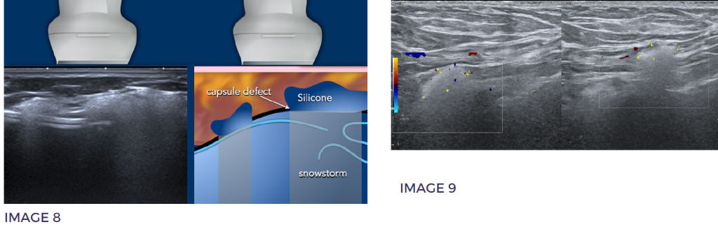
- “Snowstorm or “dirty shadowing”: acoustic shadowing caused by silicone leakage outside the capsule
- Silicone lymphadenopathy: “snowstorm” appearance of lymph nodes (Image 9)
MAMMOGRAPHY
- Popular alternative to MRI in the investigation of implant rupture as it is time and cost efficient.
- However, it is highly operator dependent and has a decrease in sensitivity in the presence of capsular contracture.
Intracapsular rupture
- Very difficult to diagnose on mammography.
- Mammography does not rule out breast implant rupture. Further investigation with Ultrasound and MRI is preferred.
Extracapsular rupture
- Loss of smooth contour of the implant is an indication of extracapsular rupture
MRI vs Ultrasound
Ultrasound is a cost effective and accessible diagnostic tool for evaluating silicone breast implant status. It can accurately diagnose silicone lymphadenopathy as an indication of gel bleed. Spit et al (2014) recommended choosing ultrasound as the initial modality in investigating breast implant rupture and only perform MRI when ultrasound results are inconclusive.
References
Szymanski KD, Mikes BA. Breast Implant Rupture. [Updated 2025 Dec 13]. In: StatPearls [Internet]. Treasure Island (FL): StatPearls Publishing; 2026 Jan-. Available from: https://www.ncbi.nlm.nih.gov/books/NBK459308/
The Radiology Assistant. (n.d.). Breast prosthesis imaging: Mammography. Retrieved from https://radiologyassistant.nl/breast/breast-prosthesis/breast-prosthesis-imaging#mammography
E. Lake, S. Ahmad, R. Dobrashian, The sonographic appearances of breast implant rupture, Clinical Radiology, Volume 68, Issue 8,2013,Pages 851-858, ISSN 0009-9260, https://doi.org/10.1016/j.crad.2013.03.014.
Soni N, Extracapsular breast implant rupture. Case study, Radiopaedia.org (Accessed on 10 Jul 2026) https://radiopaedia.org/cases/extracapsular-breast-implant-rupture-1
Spit K, Azahaf S, de Blok C …Ultrasound versus MRI for evaluation of silicone leakage from silicone breast implants, Heliyon, 2024; 10
Hillard, C., Fowler, J., Barta, R., & Cunningham, B. (2016). Silicone breast implant rupture: a review. Gland Surgery, 6(2), 163-168. doi:10.21037/gs.2016.09.1