12th October 2026, NIA Diagnostic Imaging
Contrast media play a significant role in modern medical imaging, as it helps radiologists and other healthcare professionals visualise blood vessels, organs, soft tissue, and abnormalities more clearly. It also helps in detecting lesions, assessing vascularity, and guiding treatment planning. Contrast media is generally given during specific scans such as CT, MRI, angiography, fluoroscopy, and even ultrasound.
Although contrast media are generally very safe, some patients may experience an anaphylactic reaction – similar to any other allergic reaction! Another possible complication is contrast extravasation, where contrast leaks from the vein into the surrounding tissues during injection.
Most patients receive contrast without any complications. Anaphylactic-type reactions are rare, accounting for 0.6% of cases, with only 0.04% considered aggressive (1). Almost all life-threatening contrast reactions occur within the first 20 minutes of intravenous injection (2). However, all imaging departments have protocols and emergency equipment available to manage any issues that may occur.
What is IV Contrast media?
IV contrast media are substances that temporarily change the way X-rays or other imaging tools interact with the body (3). It is a clear liquid that is injected into a vein to help with visualisation of anatomy during the scan. It does not change any internal organs and is naturally excreted by the body via urine or bowel movements. However, patients with significant kidney impairments may require additional assessment before administration of any IV contrast to ensure patient safety.
Different types of IV contrast can be administered depending on the examination:
- Iodinated Contrast – CT
Iodinated contrast is most commonly used for contrast-enhanced CT examinations and CT angiography.
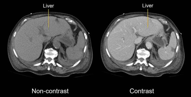
It contains iodine, which absorbs X-rays and makes blood vessels and certain organs appear more clearly on CT images. Modern iodinated contrast agents are generally non-ionic and low osmolar, which is associated with fewer adverse reactions.
2. Gadolinium-based Contrast – MRI
Gadolinium-based contrast agents (GBCAs) are used during some MRI examinations to improve visualisation of tissues, blood vessels, and areas of abnormal enhancement.
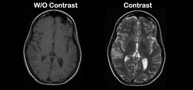
Gadolinium contrast is chemically different from iodinated contrast and is used specifically because of its properties in the MRI environment.
When IV contrast is administered, normal symptoms and sensations patients may feel include (3):
- Brief warm or flushing sensation
- Metallic taste
- Sensation of needing to urinate
- Mild nausea
These sensations usually resolve quickly and do not necessarily indicate an allergic reaction. These symptoms should be temporary and are experienced immediately after contrast is injected.
IV Contrast Reactions
Contrast reactions can range from mild symptoms to potentially life-threatening emergencies. Each reaction is categorised by severity and clinical features (4) (5) to guide management protocols.
1. Mild Reactions
Symptoms and signs include:
- Nausea
- Mild vomiting
- Transient flushing or warmth
- Mild itching
- Limited urticaria (hives)
- Persistent coughing
Management includes:
- Terminating contrast administration
- Assessment of the patient
- Observing progression of symptoms
- Escalating event if symptoms worsen
2. Moderate Reactions
Symptoms and signs include:
- More extensive urticaria
- Facial or periorbital swelling
- Persistent vomiting
- Wheezing or bronchospasm
- Significant changes in blood pressure
Management includes:
- Monitoring oxygen saturation and vital signs
- Providing oxygen where clinically indicated
- Medical review
- Administration of medications according to the department’s emergency protocol
- Continued observation
3. Severe Reactions
Symptoms and signs include:
- Anaphylaxis
- Severe bronchospasm
- Significant airway swelling
- Laryngeal oedema
- Severe hypotension
- Altered consciousness
- Cardiorespiratory arrest
Management includes:
- Calling for emergency assistance
- Maintaining airway and breathing
- Oxygen and respiratory support as clinically indicated
- Establishing or maintaining IV access
- Administration of emergency medications according to protocol
- Continuous monitoring
- CPR if required
4. Delayed Reactions
Not all reactions occur during the scan. Some hypersensitivity reactions may develop hours to
several days after contrast administration.
Possible symptoms include:
- A new skin rash
- Itching
- Redness or scattered raised lesions
Patients should contact a healthcare professional if a new or persistent rash/symptom develops after contrast administration.
Seek emergency medical assistance if there is difficulty breathing, swelling of the tongue or throat, fainting, or a widespread rash associated with blistering, skin peeling or sores affecting the mouth or eyes.
Before receiving contrast, patients should tell healthcare professionals if they have previously experienced a reaction to contrast. The imaging team can assess the previous reaction and determine the safest approach for future examinations. Depending on the circumstances, this may involve using a different contrast agent, taking additional precautions, choosing alternative imaging, or using other measures.
What is Contrast Extravasation?
Contrast extravasation occurs when IV contrast leaks out of the vein and into the surrounding soft tissues during injection. However, contrast extravasation and contrast reactions are two different reactions.
Contrast reaction is a systemic response to the contrast agent while an extravasation is contrast leaking outside the vein during the injection. Contrast extravasation can occur due to fragile or small veins, difficult venous access, cannula position, high injection pressure and flow rate.
Patients may notice common symptoms around the injection site, such as (6):
- Swelling
- Tightness or pressure
- Pain
- Redness
- Discomfort around the cannula
- Skin discolouration
Majority of extravasations are managed conservatively and resolve without long-term problems, but rare severe injuries can occur and should be promptly managed. Most extravasations are minor, but urgent assessment is important if there are signs suggesting a more significant injury.
Seek urgent medical review if there is (6) (7):
- Severe or increasing pain
- Significant or rapidly increasing swelling
- Blistering
- Skin ulceration
- Marked skin discolouration
- Numbness or altered sensation
- Reduced circulation
- Symptoms continue to worsen
What happens after a Contrast Reaction or Extravasation?
Following an event, the healthcare team may:
- Continue monitoring the patient
- Provide appropriate medical attention
- Give instructions for monitoring the affected area
- Recommend that patients tell healthcare professionals about the event before future contrast examinations
Ultimately, while IV contrast is generally safe, understanding potential complications, including contrast reactions and extravasation, is essential for both patients and healthcare professionals. Contrast enhanced imaging provides important diagnostic information that helps guide treatment and ongoing patient care.
References
1. Bell, D., & Di Muzio, B. (2016). Iodinated contrast media adverse reactions. Radiopaedia.Org. https://doi.org/10.53347/rid-42772. https://radiopaedia.org/articles/contrast-media-adverse reactions
2. American College of Radiology. (2024). ACR manual on contrast media. Acr.Org. https://www.acr.org/Clinical-Resources/Clinical-Tools-and-Reference/Contrast-Manual. https://www.acr.org/clinical-resources/clinical-tools-and-reference/contrast-manual
3. Radiological Society of North America (RSNA) and American College of Radiology (ACR). (2018). Contrast Materials. Radiologyinfo.Org. https://www.radiologyinfo.org/en/info/safety-contrast. https://www.radiologyinfo.org/en/info/safety-contrast
4. ASCIA Radiocontrast Media Hypersensitivity – Australasian Society of Clinical Immunology and Allergy (ASCIA). (n.d.). Www.Allergy.Org.Au. https://www.allergy.org.au/hp/drug-allergy/radiocontrast-media
5. Bell, D., & Di Muzio, B. (2016). Iodinated contrast media adverse reactions. Radiopaedia.Org. https://doi.org/10.53347/rid-42772. https://radiopaedia.org/articles/contrast-media-adverse-reactions
6. Radswiki. (n.d.). Contrast media extravasation | Radiology Reference Article | adiopaedia.org. Radiopaedia. https://radiopaedia.org/articles/contrast-media-extravasation.
7. Belzunegui, T., Jean Louis, C., Torrededia, L., & Oteiza, J. (2011). Extravasation of radiographic contrast material and compartment syndrome in the hand: a case report. Scandinavian Journal of Trauma, Resuscitation and Emergency Medicine, 19(1), 9. https://doi.org/10.1186/1757-7241-19
9. https://pmc.ncbi.nlm.nih.gov/articles/PMC3044092/
Figure 1: Hartung, M. P., Cadogan, M., & Cadogan, M. P. H. M. (2023, February 1). Abdominal CT: Basics. Life in the Fast Lane • LITFL. https://litfl.com/abdominal-ct-basics/ https://litfl.com/abdominal-ct-basics/
Figure 2: Naim, I. (2025, May 22). A Patient’s Guide To MRI Contrast | All You Need To Know – MRI Lucknow. MRI Lucknow. https://mrilucknow.net/mri-contrast/ https://mrilucknow.net/mri-contrast/