
25th July 2026, A/Prof Chee L Khoo
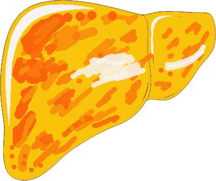
We know that 55-65% patients with type 2 diabetes (T2D) have metabolic dysfunction associated fatty liver disease (MAFLD) (1). We also know that 28% of patients with MAFLD will develop T2D if not already present by the time MAFLD is detected (2). There is emerging evidence that there are profound alterations in oral glucose handling in individuals with MAFLD even before T2D is diagnosed. Both conditions share the same pathophysiology of insulin resistance (IR) but the effects of hepatic and peripheral IR are very different. How we manage them need to be different even though both conditions are closely related.
Peripheral IR causes increased demands for insulin secretion which eventually exhaust the poor beta cells leading to eventually, insufficient insulin secretion and thence, T2D.
Hepatic IR results in ongoing glycogenolysis and gluconeogenesis leading to morning hyperglycaemia. However, while management of carbohydrate metabolism is disrupted by hepatic IR, lipid metabolism is affected differently by hepatic IR. IR leads to compensatory high circulating insulin levels and high insulin levels leads to enhanced de novo lipogenesis (DNL) triggering excessive free fatty acid (FFA) production and ultimately, elevated triglycerides (TG). The increase in FFA levels and new lipid synthesis exacerbate intrahepatic TG accumulation, leading to inflammation and oxidative stress, thereby reducing insulin sensitivity further.
It depends on how resilient the beta cells are. If there are enough tough beta cells around, MAFLD appears before T2D. But if the beta cells give up the ghost early, T2D appears before MAFLD.
There is something else that makes the whole situation between them worse: the rate of oral glucose absorption in patients. Oral glucose absorption, quantified in vivo by the rate of appearance of oral ingested glucose (RaO), is a major determinant of postprandial glycaemic and insulinaemic responses, especially in individuals without diabetes (3,4).
RaO is primarily regulated by gastric emptying, which determines the rate of entry of ingested glucose into the small intestine (5) and by the expression of glucose carriers in the upper small intestine, particularly sodium–glucose cotransporter 1 (SGLT1) (6), whose expression and activity have been associated with MAFLD risk (7-9). Rapid oral absorption leads to enhanced DNL which exacerbates MAFLD.
In a brilliant study which was published in Diabetologia recently, Trico et al looked at the kinetics of oral glucose absorption and gastric emptying, in individuals with early-stage MAFLD vs matched control individuals (10). They employed dual-tracer technique, involving both i.v. and oral administration of stable glucose isotopes, to quantify intestinal glucose absorption kinetics after an oral glucose load, while simultaneously assessing peripheral glucose disposal and hepatic glucose production. They assess gastric emptying of an OGTT assessed by the 13C-acetate breath test.
Individuals with type 2 diabetes, were taking medications influencing glucose or lipid metabolism, or were pregnant or lactating were excluded. MAFLD was defined as the presence of steatotic liver disease confirmed on ultrasound, associated with at least one cardiometabolic risk factor, in the absence of significant alcohol consumption (>140 g/week or >20 g/day for female participants; >210 g/week or >30 g/day for male participants) and other discernible causes of liver disease.
Plasma glucose concentrations at fasting and 2 h post-OGTT were similar between the two groups but the RaO was substantially higher in the MAFLD group in the first hour of the OGTT. In the MAFLD group compared with the control group, the total amount of absorbed oral glucose was 51.6% higher at 1 h during the OGTT (+6.4±1.8 g, p=0.001) and 53.2% higher at 2 h (+10.7±3.6 g, p=0.005). Results were materially unchanged in BMI-matched sensitivity analyses.
Plasma insulin levels were higher in the MAFLD group than the control group during fasting (+38±10 pmol/l, p=0.0004) and throughout the OGTT. Oral glucose absorption was identified as a significant predictor of MAFLD.
After the glucose drink, the blood glucose response was higher in the group with HSI ≥36 (group effect: p=0.010) (Fig. 5a); this group showed higher blood glucose levels at 1 h and 2 h post-OGTT than the group with HSI <30. This is not due to a more rapid gastric emptying time. Between these two groups, there were no differences in T50 or gastric retention (expressed as % of baseline) at 1 h post-OGTT suggesting that the higher glucose response is not related to a more rapid gastric emptying.
Glucose absorbed from the gut is preferentially delivered to the liver via the portal vein, where excess glucose influx can stimulate triglyceride accumulation and promote MAFLD progression from simple steatosis to steatohepatitis and fibrosis. Additionally, increased oral glucose absorption translates into higher plasma glucose excursions and sustained hyperinsulinaemia, which may further stimulate DNL and impair lipid export from the liver (11).
Now, the pathogenesis of MASLD is multifactorial and intrinsically linked to metabolic derangements, with chronic hyperglycaemia, insulin resistance and beta cell dysfunction recognised as key drivers of hepatic lipid accumulation and inflammation. As you can see, all those risk conditions feeds off each other. Since MAFLD is so common amongst patients who are overweight or obese and in patients with diabetes, we need to confirm the diagnosis with ultrasound and not rely on abnormal liver functions tests.
Further, knowing that patients with MAFLD have abnormal capacity to handle glucose, it is vital that we reduce their carbohydrate intake right from the outset. Low carbohydrate diet as part of an overall balance diet is in the International Multidisciplinary Expert Consensus Statement (2024) (12).
References:
- Younossi Z, Golabi P, Price J. et al. The Global Epidemiology of Nonalcoholic Fatty Liver Disease and Nonalcoholic Steatohepatitis Among Patients With Type 2 Diabetes. Clinical Gastroenterology and Hepatology, 2024; 22, 1999-2010.e8
- Cao L, An Y, Liu H, Jiang J, Liu W, Zhou Y, Shi M, Dai W, Lv Y, Zhao Y, Lu Y, Chen L, Xia Y. Global epidemiology of type 2 diabetes in patients with NAFLD or MAFLD: a systematic review and meta-analysis. BMC Med. 2024 Mar 6;22(1):101
- Trico D, Galderisi A, Mari A, Santoro N, Caprio S (2019) One-hour post-load plasma glucose predicts progression to prediabetes in a multi-ethnic cohort of obese youths. Diabetes Obes Metab 21(5):1191–1198
- Phillips LK, Deane AM, Jones KL, Rayner CK, Horowitz M (2015) Gastric emptying and glycaemia in health and diabetes mellitus. Nat Rev Endocrinol 11(2):112–128.
- Wu T, Rayner CK, Jones KL, Xie C, Marathe C, Horowitz M (2020) Role of intestinal glucose absorption in glucose tolerance. Curr Opin Pharmacol 55:116–124.
- Wright EM, Loo DD, Hirayama BA (2011) Biology of human sodium glucose transporters. Physiol Rev 91(2):733–794.
- Fiorentino TV, De Vito F, Suraci E et al (2022) Augmented duodenal levels of sodium/glucose co-transporter 1 are associated with higher risk of nonalcoholic fatty liver disease and noninvasive index of liver fibrosis. Diabetes Res Clin Pract 185:109789.
- Dobbie LJ, Cuthbertson DJ, Hydes TJ, Alam U, Zhao SS (2023) Mendelian randomisation reveals sodium-glucose cotransporter-1 inhibition’s potential in reducing non-alcoholic fatty liver disease risk. Eur J Endocrinol 188(6):K33–K37.
- Honda Y, Ozaki A, Iwaki M et al (2021) Protective effect of SGL5213, a potent intestinal sodium-glucose cotransporter 1 inhibitor, in nonalcoholic fatty liver disease in mice. J Pharmacol Sci 147(2):176–183
- Tricò, D., Wu, T., Cimbalo, N. et al. Oral glucose absorption is enhanced in early metabolic dysfunction-associated steatotic liver disease. Diabetologia 69, 2073–2087 (2026)
- Loomba R, Friedman SL, Shulman GI (2021) Mechanisms and disease consequences of nonalcoholic fatty liver disease. Cell 184(10):2537–2564.
- Zeng XF, Varady KA, Wang XD, et al. The role of dietary modification in the prevention and management of metabolic dysfunction-associated fatty liver disease: An international multidisciplinary expert consensus. Metabolism. 2024 Dec;161:156028