
3rd January 2026, A/Prof Chee L Khoo
Women, in general, have a lower cardiovascular (CV) risk because of the “protection” from the oestrogen exposure from puberty onwards. Of course, that “protection” ends when a woman becomes menopausal and indeed, the prevalence of CV events very quickly approaches those of men within a few years. One of the most difficult decisions to make is when to treat elevated lipids in women. In addition to the common CV risk factors that affect men, we looked at adverse pregnancy outcomes (APO) at GPVoice in February 2025. New data from a large US population-based cohort study highlighted uterine fibroids as a potential signal of increased long-term risk for atherosclerotic cardiovascular disease (ASCVD).
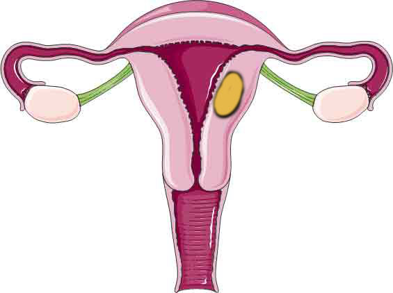
Uterine leiomyomas (or fibroids) are the most common benign neoplasm of the female reproductive tract, present in up to 77% of reproductive-age women (1,2). The development of fibroids would appear to be a disease process intrinsic to the uterus but there is accumulating evidence which suggests that fibroid development may be linked with the systemic vasculature system.
Fibroids actually share many pathogenic features with ASCVD. Both originate from monoclonal smooth muscle cells, and fibroid growth involves smooth muscle proliferation, fibrosis, and calcification. This process is similar to vascular remodelling disorders (3-6). In addition, fibroids can also release inflammatory cytokines (eg, interleukin‐6, tumour necrosis factor‐α) and chemokines within the uterine environment (3, 7). Like all inflammatory cytokines, some of these chemicals may enter systemic circulation, causing generalised systemic inflammation and oxidative stress, thereby adding to the ASCVD risk.
This pathologic link is not new. Hypertensive disorders and preeclampsia has been positively associated with fibroid occurrence (8,9). Further, polymorphisms in genes regulating vascular tone and arterial blood flow including the angiotensin converting enzyme (ACE) and angiotensin receptor have been associated with fibroids, also suggesting a possible underlying pathological link (10,11).
Lauglin-Tomaso et al reported in a study of 972 women from the Coronary Artery Risk Development in Young Adults (CARDIA) study cohort (12). Women 35–49 years were screened for the uterine fibroids and followed for up to 25 years. Most CVD risk factors were more common in women with fibroids but these differences were not statistically significant after adjusting for confounders, including blood pressure and body mass index, likely due to limited power for rare outcomes. Further, presence of fibroids was not associated with subclinical CVD.
DiTosto J et al recently reported on a study from a US population-based cohort. 450 177 women with fibroids were compared with 2 250 885 controls without fibroids (13). They were followed up for about 5 years. Incident ASCVD, a composite of coronary artery disease, cerebrovascular disease, and peripheral artery disease, was evaluated. Secondary outcomes examined individual ASCVD components of coronary artery disease, cerebrovascular disease, and peripheral artery disease.
Individuals with fibroids were exact age‐matched to 5 controls with no prior fibroid claim but with a claim for a general gynaecological examination within 30 days of the matched fibroid date. Matched controls were eligible to be re‐recruited as exposed if later diagnosed with fibroids.
The average age at baseline of 40.93 years. Average follow‐up time was 5.18 years in the fibroid group and 4.02 years in the control group. Before weighting, the proportion of Black individuals and the prevalence of ASCVD risk factors were higher in the fibroid group.
At the 1 year mark, there was an increased ASCVD risk in the fibroid group (1‐year risk ratio: 2.47; 1‐year risk difference, 0.41%). At the 10-year mark, the risk ratio was 1.81 with the 10‐year risk difference of 2.40%. The increased risk was consistent for all individual components of ASCVD.
Blood pressure and body mass index were not available in this study but they adjusted for ASCVD risk factors using both diagnoses and medications on file. Mild hypertension or slight elevated BMI may not be captured. The increased ASCVD risk associated with fibroids remained even after accounting for these factors.
The latest study by DiTosto J et al is a much bigger study involving 2.7 milllion patients. Women with fibroids are 1.8 times more likely to developed ASCVD compared with women who did not have fibroid. What it is telling us is that when we diagnosed fibroids in women, whether symptomatic or not, it should prompt us to re-evaluate the CV risk profile of the woman in front of us. We already have a large number of CV risk enhancers which influence our CV risk assessment in women and fibroids represent another risk factor to consider.
References
- Cramer SF, Patel A. The frequency of uterine leiomyomas. Am J Clin Pathol. 1990 Oct;94(4):435-8.
- Okolo S. Incidence, aetiology and epidemiology of uterine fibroids. Best Pract Res Clin Obstet Gynaecol. 2008 Aug;22(4):571-88.
- Kirschen GW, AlAshqar A, Miyashita‐Ishiwata M, Reschke L, El Sabeh M, Borahay MA. Vascular biology of uterine fibroids: connecting fibroids and vascular disorders. Reproduction. 2021;162:R1–r18.
- Silver MA, Raghuvir R, Fedirko B, Elser D. Systemic hypertension among women with uterine leiomyomata: potential final common pathways of target end‐organ remodeling. J Clin Hypertens (Greenwich). 2005;7:664–668
- Moss NS, Benditt EP. Human atherosclerotic plaque cells and leiomyoma cells. Comparison of in vitro growth characteristics. Am J Pathol. 1975;78:175–190
- Malik M, Norian J, McCarthy‐Keith D, Britten J, Catherino WH. Why leiomyomas are called fibroids: the central role of extracellular matrix in symptomatic women. Semin Reprod Med. 2010;28:169–179
- AlAshqar A, Patzkowsky K, Afrin S, Wild R, Taylor HS, Borahay MA. Cardiometabolic risk factors and benign gynecologic disorders. Obstet Gynecol Surv. 2019;74:661–673
- Pan L, Fu Z, Yin P, Chen D. Pre-existing medical disorders as risk factors for preeclampsia: an exploratory case-control study. Hypertens Pregnancy. 2019 Nov;38(4):245-251.
- Roberts WE, Fulp KS, Morrison JC, Martin JN Jr. The impact of leiomyomas on pregnancy. Aust N Z J Obstet Gynaecol. 1999 Feb;39(1):43-7.
- Keshavarzi F, Teimoori B, Farzaneh F, Mokhtari M, Najafi D, Salimi S. Association of ACE I/D and AGTR1 A1166C Gene Polymorphisms and Risk of Uterine Leiomyoma: A Case-Control Study. Asian Pac J Cancer Prev. 2019 Sep 1;20(9):2595-2599.
- GOMAA SW, ZAKI AM, EL-ATTAR EA, et al. Polymorphisms of Renin Angiotensin System Genes in Uterine Leiomyomas Among Egyptian Females. Journal of Clinical Gynecology & Obstetrics, 4, 170–176
- Laughlin-Tommaso SK, Fuchs EL, Wellons MF, Lewis CE, Calderon-Margalit R, Stewart EA, Schreiner PJ. Uterine Fibroids and the Risk of Cardiovascular Disease in the Coronary Artery Risk Development in Young Adult Women’s Study. J Womens Health (Larchmt). 2019 Jan;28(1):46-52.
- DiTosto JD, Lewey J, Zee J, et al. Association Between Uterine Fibroids and Risk of Atherosclerotic Cardiovascular Disease. J Am Heart Assoc. 2025 Dec 10:e044014.