
12th August 2022, Dr Chee L Khoo
We used to think of the islets of Langerhans as tenants in the exocrine pancreas boarding house. Like two separate cell types living under the same roof – one endocrine and the other exocrine. Both cell types happen to be involved in the business of nutrient digestion but they perform totally separate functions. Patients with diabetes are more likely to get pancreatic cancers and pancreatic exocrine insufficiency. On the other hand, patients with pancreatic disease are more likely to develop diabetes mellitus (DM) too. Thus, the two cell types are more closely related than we first thought. Most interesting. We should pursue this relationship a bit more.
Type 3c diabetes also called diabetes of the exocrine pancreas (DEP). It includes glucose dysmetabolism secondary to disorders of the exocrine pancreas. It is also referred to as pancreatogenic diabetes. It is reported to account for up to 9% of patients hospitalised with diabetes. T3c is frequently misdiagnosed as T2D.
75% of DEP is caused by chronic pancreatitis. The other causes of DEP include acute pancreatitis, cystic fibrosis, pancreatic adenocarcinoma, pancreatic surgical resections and autoimmune pancreatitis. Less common causes of DEP are hereditary hemochromatosis, transfusion overload, and thalassemia major.
Chronic pancreatitis (CP)
You would think that diabetes develops because chronic pancreatitis have destroyed the house which the islet of Langerhans cells live in and as a result, there are fewer beta cells. Insulin secretion is then reduced and diabetes develops. It is a little more complicated than that. The pathway for progression to diabetes is a quantitative reduction in beta cells as well as a qualitative reduction in beta cell function.
There is gradual beta cell loss with intra-pancreatic inflammation. The inflammation is coordinated by reduction in T-helper 1 and T-helper 17 cells and increase in interferon-γ. Ongoing inflammation leads to beta cell death via apoptosis. This is the quantitative reduction of beta cells.
There is further reduction in cell numbers comes from beta cell dedifferentiation. Under certain metabolic stress conditions, such as oxidative stress, endoplasmic reticulum (ER) stress, inflammation, hypoxia, or even glucotoxicity induced by hyperglycaemia, mature β-cells lose their phenotype and regress to a precursor-like dedifferentiated state [1]. Through dedifferentiation, the β-cells lose the genes that are responsible for their identity and maturity and acquire a new progenitor-like phenotype with a different potency [2]. The degree of dedifferentiation was correlated with the severity of the inflammation, fibrosis, or atrophy of the pancreas [1].
To make matters worse, long before the beta cell numbers drop, alteration of the internal pancreatic environment affects islet cell secretory function. Proinflammatory cytokines, such as interleukin 1b (IL-1b), tumour necrosis factor α (TNF-a), and IFN-γ, play a role in inhibiting the hormone secretion activity [3]. Insulin-like growth factor-1 (IGF-1) and gastrointestinal hormones, together with cytokines and chemokines, have a significant role in influencing the islet function. Low concentrations of IL-1b stimulate β-cell proliferation, while higher concentrations lead to functional impairment and β-cell loss through necrosis and cell apoptosis. The inflammatory cytokines also inhibit GLP1 and GIP receptors.
DM is a delayed complication of long-standing CP. In one study, approximately 50% of patients progress to DM 10 years after a diagnosis of CP and 83% at 25 years after diagnosis [4]. Similarly, Pan et al. reported that 90% of patients had diabetes of the exocrine pancreas after 50 years of CP evolution [5]. But Wang et al. in a study on a Chinese population, with found a cumulative rate of 24% at 10 years and of 52% at 20 years of CP [6]. DM can develop earlier in the course of CP. In a study by Olesen at al., 61% of patients developed DM less than 5 years after the diagnosis of CP [7].
The general risk factors for developing diabetes in patients with CP are dyslipidaemia, obesity, age at diagnosis of CP, duration of CP, presence of pancreatic calcification, degree of pancreatic resection and presence of pancreatic exocrine insufficiency (7).
Acute Pancreatitis
One of the most relevant causes of DM after AP is the loss of beta cells from pancreatic tissue loss. Extensive pancreatic necrosis determines the loss of functional pancreatic tissue and reduction of beta -cells [8]. Some authors did not find the relationship between the severity of pancreatitis and the development of diabetes (9). Perhaps, AP triggers an immune response in genetically susceptible patients for developing DM [9].
Another dominant role in developing T3cDM after AP could be the insulin resistance [8]. Once again, pancreatic inflammation is involved but this time, the dominant cytokine IL-6 causes impaired phosphorylation of the insulin receptor and insulin receptor substrate-1 and leading to insulin resistance. Increased lipolysis leads to increased glycerol and triglycerides which leads to an increase in IL-6.
The relationship between AP and DM cuts both ways. Patients with DM are more likely to get AP. A population-based cohort study in Taiwan in 2016 [10] described the bidirectional relationship between AP and DM. The adjusted hazard ratio of AP was significantly higher in patients with DM, with higher values in those with a history of hyperglycaemic crisis episodes, including ketoacidosis and a hyperosmolar hyperglycaemic. Coming from the other direction, adjusted hazard ratio of DM was increased in patients with AP compared to the general population.
Cystic Fibrosis (CF)
Cystic fibrosis (CF), an autosomal recessive disease, is a multisystemic disorder caused by abnormal chloride and bicarbonate transport related to mutations of the CFTR (cystic fibrosis transmembrane conductance regulator) protein, which leads to thick secretions in the lung, pancreas, liver, intestine, and reproductive tract. Aside from the well-known exocrine manifestations, CF may be responsible for alterations in the pancreatic endocrine function and glucose metabolism. CF-related diabetes (CFRD) affects approximately 35% of adults with CF [32]. It is considered a form of T3cDM, but is often recognized as a distinct item, as it has features of both T1DM and T2DM.
It is thought that the development of DM in CF stems primarily from beta-cell loss and intra-islet inflammation with amyloid deposition. Patients with CFRD have a decreased density of islet tissue compared with CF patients without diabetes [11-14]. The aetiology of beta cell loss is multiple: autodigestion from pro-enzyme retention in the pancreatic ducts, oxidative stress of beta cells and the malabsorption of dietary antioxidants in CF subjects, together with the misfolded CFTR proteins in the ER of the beta-cells, can induce ER stress and beta-cell apoptosis [13]. Hyperglycaemia can add further insult leading to further apoptosis.
Once again, pancreatic inflammation involving IL-6, TH1, TH17 is present but islet amyloid is also involved in early damage to the beta cells. Another responsible mechanism could be the CFTR expression in pancreatic endocrine cells. Bellin et al. revealed that the CFTR is directly implicated in insulin secretion, and the correction of CFTR function could be the key to the improvement of glucose tolerance in CF patients [15]. accumulation of misfolded CFTR proteins in the endoplasmic reticulum (ER) of the beta-cells as a determinant of beta-cell apoptosis [13].
Pancreatic surgery
The part of the resected pancreas plays an important role, as pancreatic endocrine cells are distributed non-uniformly along the gland, with beta-cells mostly in the tail, pancreatic polypeptide cells in the head and beta-cells throughout the entire pancreas. The relationship between the development of DM and the extent of pancreatic resection is conflicting. The site of resection seems to affect insulin sensitivity. Jun Ishida et al., it was found that glucose tolerance and insulin sensibility are better after pancreaticoduodenectomy compared to distal pancreatectomy [16] whereas pancreatoduodenectomy was correlated with a significantly higher incidence of diabetes compared to medical treatment (17). King et al. found a minimal (9%) rate of new onset diabetes (NOD) after distal pancreatectomy (18).
Beger et al. [19] showed an increased risk of postoperative NOD and pancreatic exocrine insufficiency in patients with pancreatoduodenectomy when compared to duodenum-preserving pancreatic head resection for benign tumours (NOD in 15% of patients with pancreato-duodenectomy vs. 6% in patients with preserved duodenum in the meta-analysis). Singh et al. [20] found that a percentage of <48.8% of residual pancreatic volume could be a significant risk factor for impaired glucose tolerance and progression to DM [20]. It is thought that the reduction in pancreatic endocrine and exocrine secretion is more of a consequence of duodenal removal, as a major regulator of hormone secretion, and less of a consequence of pancreatic head removal [19].
Surprisingly, there are cases where pancreatoduodenectomy may improve the glucose metabolism. In patients with pancreatic cancer, pancreatoduodenectomy may improve glycaemic control through the decreased insulin resistance. A better effect is seen in patients with NOD than in those with long-standing DM.
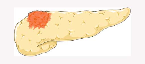
We recently discussed the relationship between pancreatic adenocarcinoma and DM on GPVoice.
In summary, the mechanism involved in the development of diabetes related to pancreatic diseases, be it pancreatitis, pancreatic cancer, cystic fibrosis or pancreatic surgery is not just related to pancreatic inflammation causing reduced beta cells but is also related to impairment in beta cell function leading to reduce insulin secretion. Screening for glucose metabolism impairment and DM has to be routine after flares of pancreatic exocrine diseases or in established diseases of the exocrine pancreas, not only at the moment of diagnosis, but also in the long-term follow-up.
References:
- Sun, J, Ni, Q, Xie, J. et al J. Clin. Endocrinol. Metab. 2019, 104, 83–94.
- Bensellam, M, Jonas, J-C.; Laybutt, D.R. J. Endocrinol. 2018, 236,
- Butler, A.E.; Janson, J.; Bonner-Weir, S et al. Diabetes 2003, 52, 102–110
- Malka, D.; Hammel, P.; Sauvanet, A.; et al. Gastroenterology 2000, 119, 1324–1332
- Pan, J.; Xin, L.; Wang, D.; et al. Medicine 2016, 95, e3251
- Wang, W.; Guo, Y.; Liao, Z. Pancreas 2011, 40, 206–212.
- Olesen, S.S.; Poulsen, J.L.; Novovic, S. United Eur. Gastroenterol. J. 2020, 8, 453–461
- Tu, J.; Yang, Y.; Zhang, J. Medicine 2018, 97, e10713.
- Das, S.L.M.; Singh, P.P.; Phillips, A.R.J.; et al. Gut 2014, 63, 818–831
- Lee, Y.-K.; Huang, M.-Y.; Hsu, C.-Y.; Su, Y.-C. Medicine 2016, 95, e2448
- Hart, N.; Aramandla, R.; Poffenberger, G. JCI Insight. 2018, 3, e98240
- Hull, R.L.; Gibson, R.L.; McNamara, S. Diabetes Care 2018, 41, 823–830.
- Barrio, R. Eur. J. Endocrinol. 2015, 172, R131–R141.
- Hameed, S.; Jaffé, A.; Verge, C.F. Pediatr. Pulmonol. 2011, 46, 747–760
- Bellin, M.D.; Laguna, T.; Leschyshyn, J. Pediatr. Diabetes 2013, 14, 417–421
- Ishida, J.; Toyama, H.; Matsumoto, I. J. Am. Coll. Surg. 2021, 233, 753–762
- Balduzzi, A.; Marchegiani, G.; Andrianello, S. Pancreatology 2020, 20, 193–198
- King, J.; Kazanjian, K.; Matsumoto, J. J. Gastrointest. Surg. 2008, 12, 1548–1553
- Beger, H.G.; Mayer, B.; Poch, B. HPB 2020, 22, 809–820
- Singh, A.N.; Pal, S.; Kilambi, R. J. Surg. Res. 2018, 227, 211–219
- Marina Ciochina, Daniel Vasile Balaban, George Manucu. Biomolecules 2022, 12, 618.